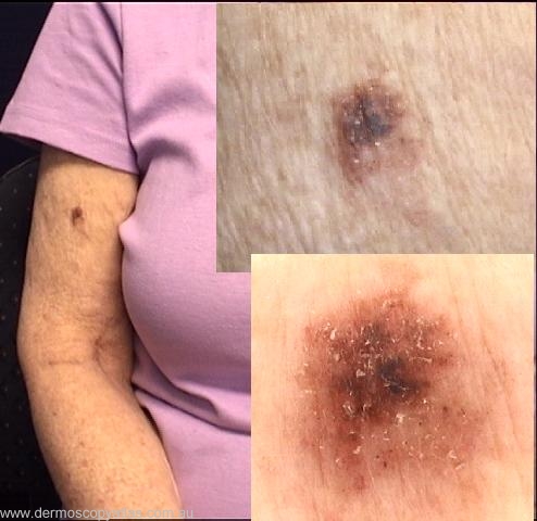
Loading
TeleDermConsult-logo
  • Recent Submissions
  • Search
  • Diagnosis
  • Quiz
  • Image comparison
  • Submit
  • Teaching Cases
  • Index
  • About

Teaching Cases


teach1

History

This 65 yr old female lives in Sweden, and was visiting her daughter after several years. The lesion on her arm is long standing but her daughter felt it was larger and darker.

teach1

View Full Size Image

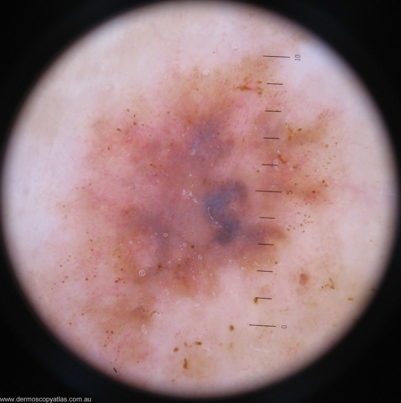
Question: What is your clinical diagnosis?

Answer: The clinical could be a seborrhoeic keratosis with the rough surface but the varying colours are a bit worrying.


Question: What is your dermatoscopic diagnosis?

Answer: The dermoscopy shows multiple peripheral dots with a blue grey veil centrally and a variable network. Vessels are not easily visible. There are no features to support a diagnosis of seborrhoeic keratosis.


Question: What do you think the histology will show?

Answer: Histology showed LEVEL 4 MALIGNANT MELANOMA, BRESLOW THICKNESS 1.37MM. SUPERFICIAL SPREADING INTRAEPIDERMAL COMPONENT.


Next Question
  • Recent Submissions
  • Search
  • Diagnosis
  • Quiz
  • Image comparison
  • Submit an Image
  • Teaching Cases
  • Index
  • About

Copyright 2022. Dermoscopy Atlas. ALL Rights Reserved.

Change Image