Loading
TeleDermConsult-logo
  • Recent Submissions
  • Search
  • Diagnosis
  • Quiz
  • Image comparison
  • Submit
  • Teaching Cases
  • Index
  • About

Teaching Cases


teach1

History

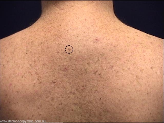
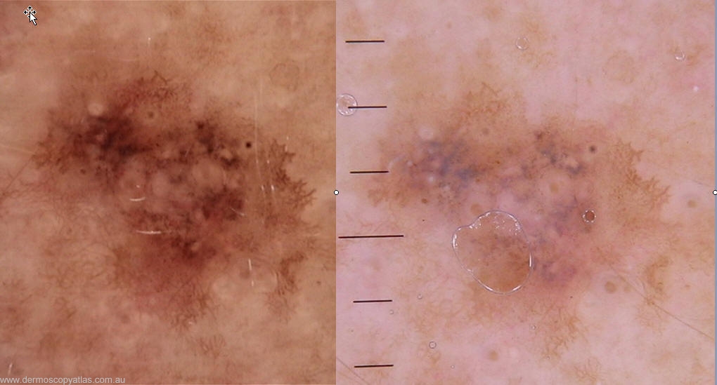
This 34 year old male presented for a routine skin check, and this more prominant "freckle" was noted on his upper back. The image on the left is mole max and on the right heine.

teach1

View Full Size Image

Question: What is your clinical diagnosis?

Answer: A more prominent freckle, Lentigo maligna or more florid melanoma in situ.


Question: What is your dermatoscopic diagnosis?

Answer: Grey dots of regression without a prominent network. The regression is worrying. You need to biopsy to see the underlying lesion.


Question: What do you think the histology will show?

Answer: Histology - Sections show a solar lentigo. There is patchy inflammation and surface parakeratosis. Some loss of melanin into the underlying papillary dermis is noted. No significant melanocytic atypia or hyperplasia is evident. There is no evidence of mal


Next Question
  • Recent Submissions
  • Search
  • Diagnosis
  • Quiz
  • Image comparison
  • Submit an Image
  • Teaching Cases
  • Index
  • About

Copyright 2022. Dermoscopy Atlas. ALL Rights Reserved.

Change Image