Loading
TeleDermConsult-logo
  • Recent Submissions
  • Search
  • Diagnosis
  • Quiz
  • Image comparison
  • Submit
  • Teaching Cases
  • Index
  • About

Teaching Cases


teach1

History

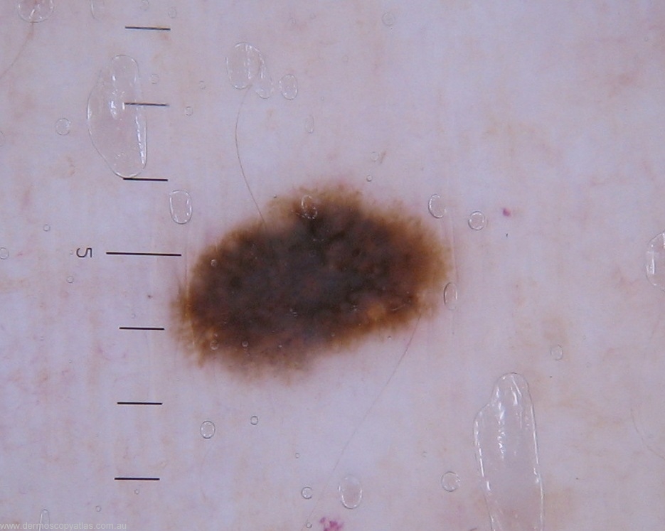
This 58 year old female presented for a routine skin check, this lesion over the left biceps had been present as long as she could recall. The lesion was monitored and due for review at 3 months however the patient returned at 6 months and indicated the lesion was getting darker. The mole max comparison is shown below after 6 months with the Heine image also at 6 months review.

teach1

View Full Size Image

teach1

View Full Size Image

Question: What is your clinical diagnosis?

Answer: Difficult on this image. The history suggests a benign lesion but it is very dark and patients sometimes mislead!


Question: What is your dermatoscopic diagnosis?

Answer: The review molemax images show a change . The Heine gives more detail showing a symmetrical pimented lesion suggesting a dysplastic nevus.


Question: What do you think the histology will show?

Answer: Histology showed a severely dysplastic junctional naevus. No dermal or epidermal invasion is seen.


Next Question
  • Recent Submissions
  • Search
  • Diagnosis
  • Quiz
  • Image comparison
  • Submit an Image
  • Teaching Cases
  • Index
  • About

Copyright 2022. Dermoscopy Atlas. ALL Rights Reserved.

Change Image