Loading
TeleDermConsult-logo
  • Recent Submissions
  • Search
  • Diagnosis
  • Quiz
  • Image comparison
  • Submit
  • Teaching Cases
  • Index
  • About

Teaching Cases


teach1

History

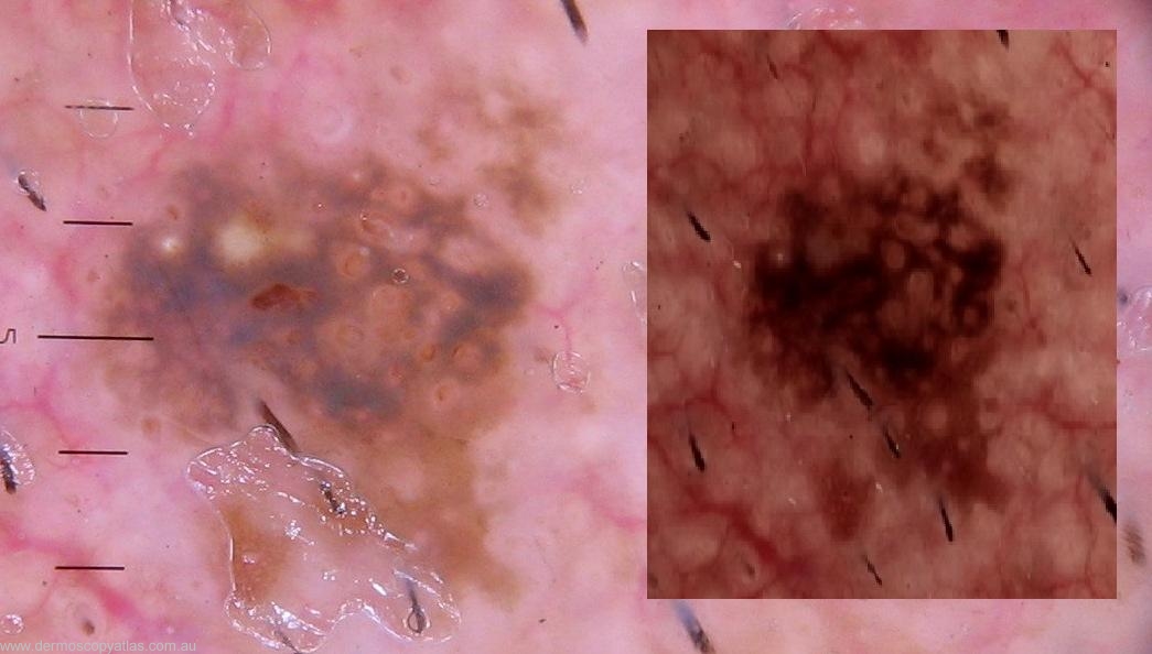
This 66 year old male construction worker presented with several BCC, this R preauricular lesion was also noted. Consider Solar lentigo, Pigmented solar keratosis,Seborrhoeic keratosis, Melanoma in situ or Melanoma.

teach1

View Full Size Image

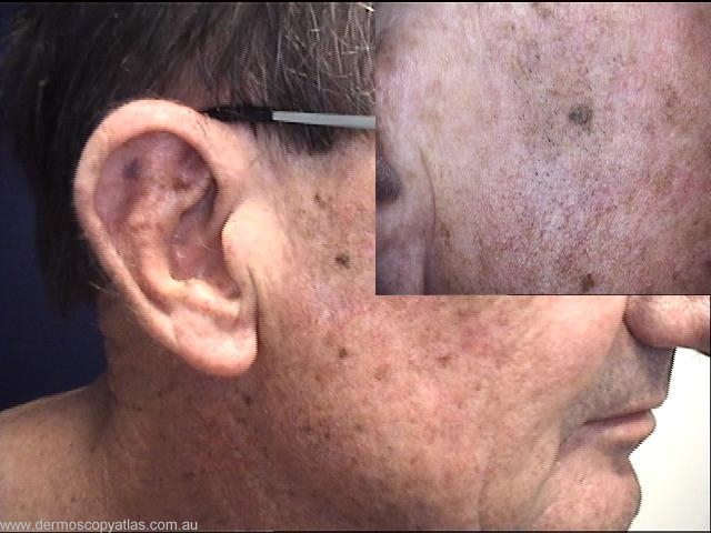
Question: What is your clinical diagnosis?

Answer: Clinically you have a background macular pigmented area with a bron papule on it. This looks like a seborrhoeic keratosis.


Question: What is your dermatoscopic diagnosis?

Answer: This shows a facial pseudonetwork with the opening of hair follicles and some white milia. The background pigmentation does not show a network. This is a seborrhoeic keratosis dermatoscopically.


Question: What do you think the histology will show?

Answer: Histology showed an actinic pattern seborrhoeic keratosis


Next Question
  • Recent Submissions
  • Search
  • Diagnosis
  • Quiz
  • Image comparison
  • Submit an Image
  • Teaching Cases
  • Index
  • About

Copyright 2022. Dermoscopy Atlas. ALL Rights Reserved.

Change Image