
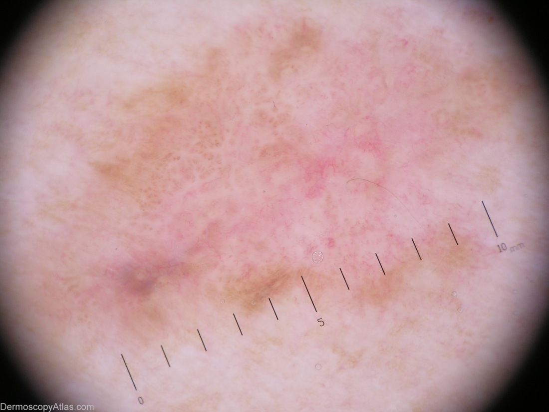
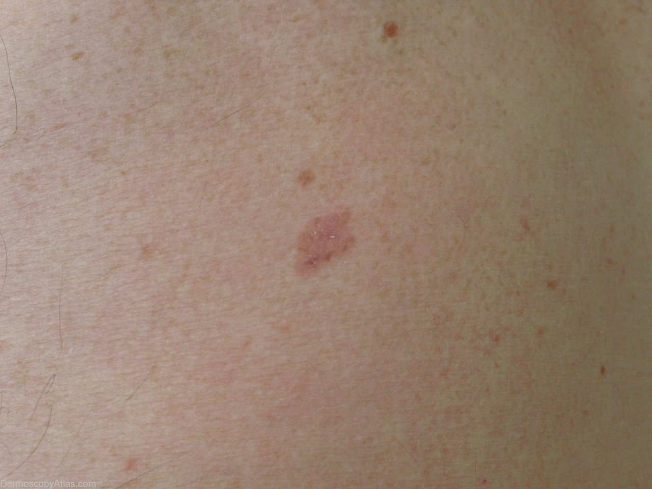
Site: Back
Diagnosis: Melanoma amelanotic
Sex: M
Age: 70
Type: Heine
Submitted By: Jean-Yves Gourhant
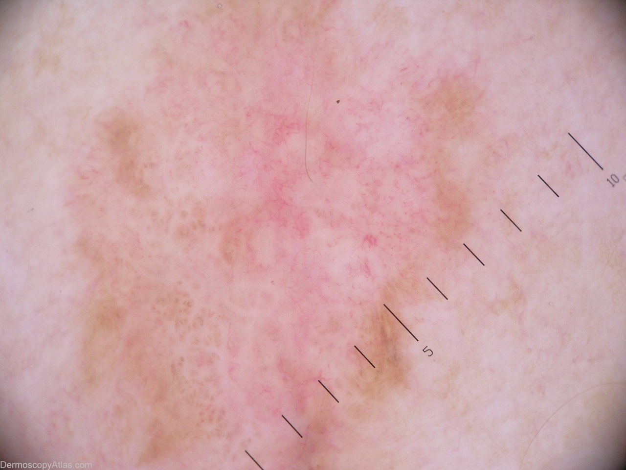
Description: The lesion shows mostly vessels, which fit neither with a diagnosis of seborrheic keratosis, nor with a basal cell carcinoma: mainly dotted vessels, and irregular linear vessels. There also white areas of regression, and an inversed network. It is circled by a brown edge, but it is difficult to find any sign in favour of a network. Finally, a blue-grey clod was a pitfall, in favour a basal-cell carcinoma.
History: This lesion was discovered by a systematic exam. Clinically it was not much concerning, but as the dermoscopic view showed no clear (benign) sign, excision became mandatory. The pathology revealed a compound melanocytic proliferation, with a strong and atypical junctional activity. The final diagnosis being Melanoma in situ, which developed from a compound nevus.