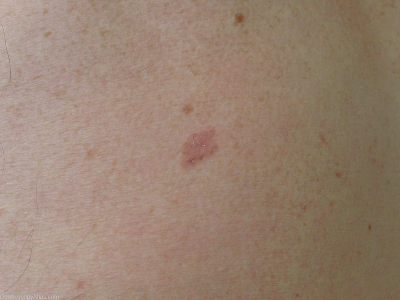
Loading
TeleDermConsult-logo
  • Recent Submissions
  • Search
  • Diagnosis
  • Quiz
  • Image comparison
  • Submit
  • Teaching Cases
  • Index
  • About

<< Back to Diagnosis Page

Image Number #2902 (Melanoma amelanotic)

Site: Back

Diagnosis: Melanoma amelanotic

Sex: M

Age: 70

Type: Heine

Submitted By: Jean-Yves Gourhant

View Full Size Image

Click Here to View Details

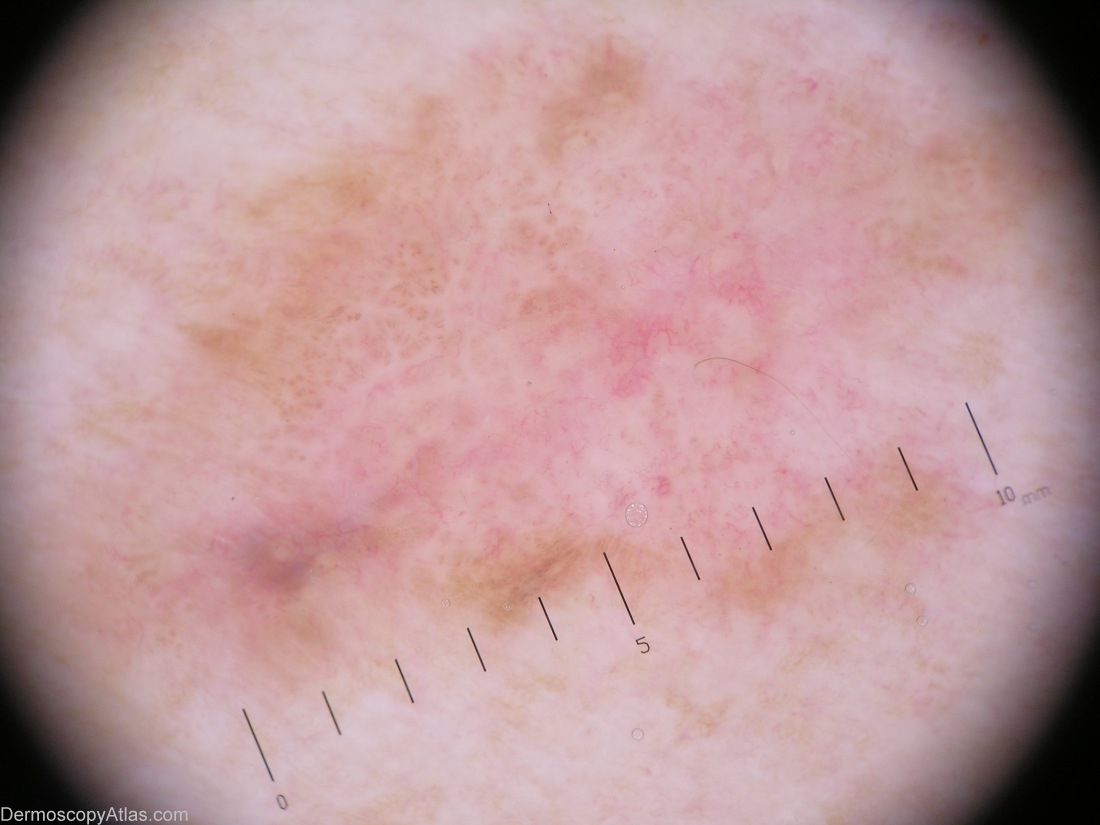
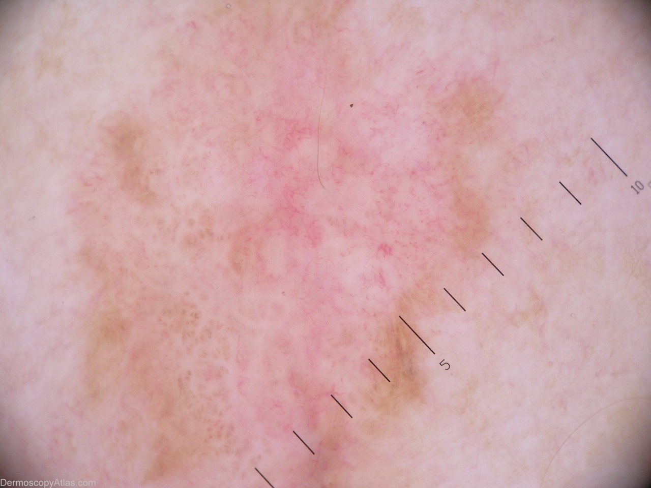
Description: An inconspicuous lesion of the back: mainly pink, a bit scaly.

History: This lesion was discovered by a systematic exam. Clinically it was not much concerning, but as the dermoscopic view showed no clear (benign) sign, excision became mandatory. The pathology revealed a compound melanocytic proliferation, with a strong and atypical junctional activity. The final diagnosis being Melanoma in situ, which developed from a compound nevus.

DermNetNZ   eMedicine   PubMed   Dermatology Online   Archives   JAAD for "Melanoma amelanotic"

  • Recent Submissions
  • Search
  • Diagnosis
  • Quiz
  • Image comparison
  • Submit an Image
  • Teaching Cases
  • Index
  • About

Copyright 2022. Dermoscopy Atlas. ALL Rights Reserved.

Change Image