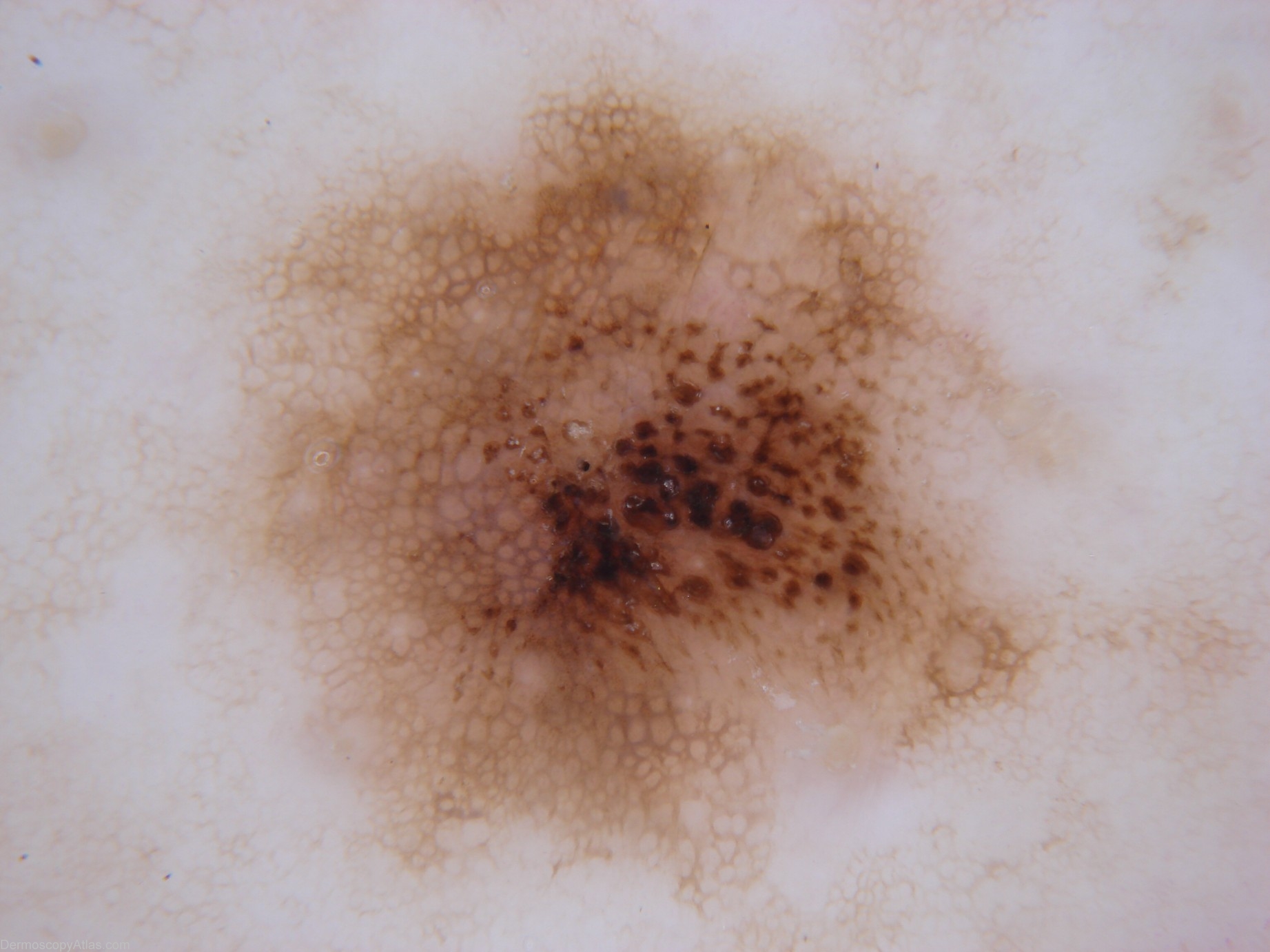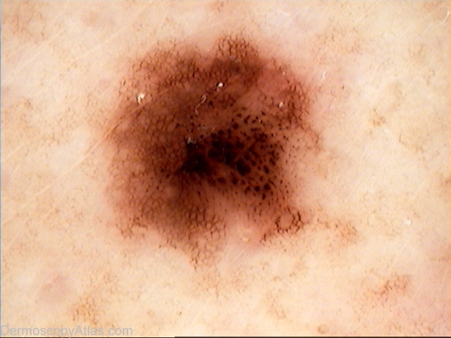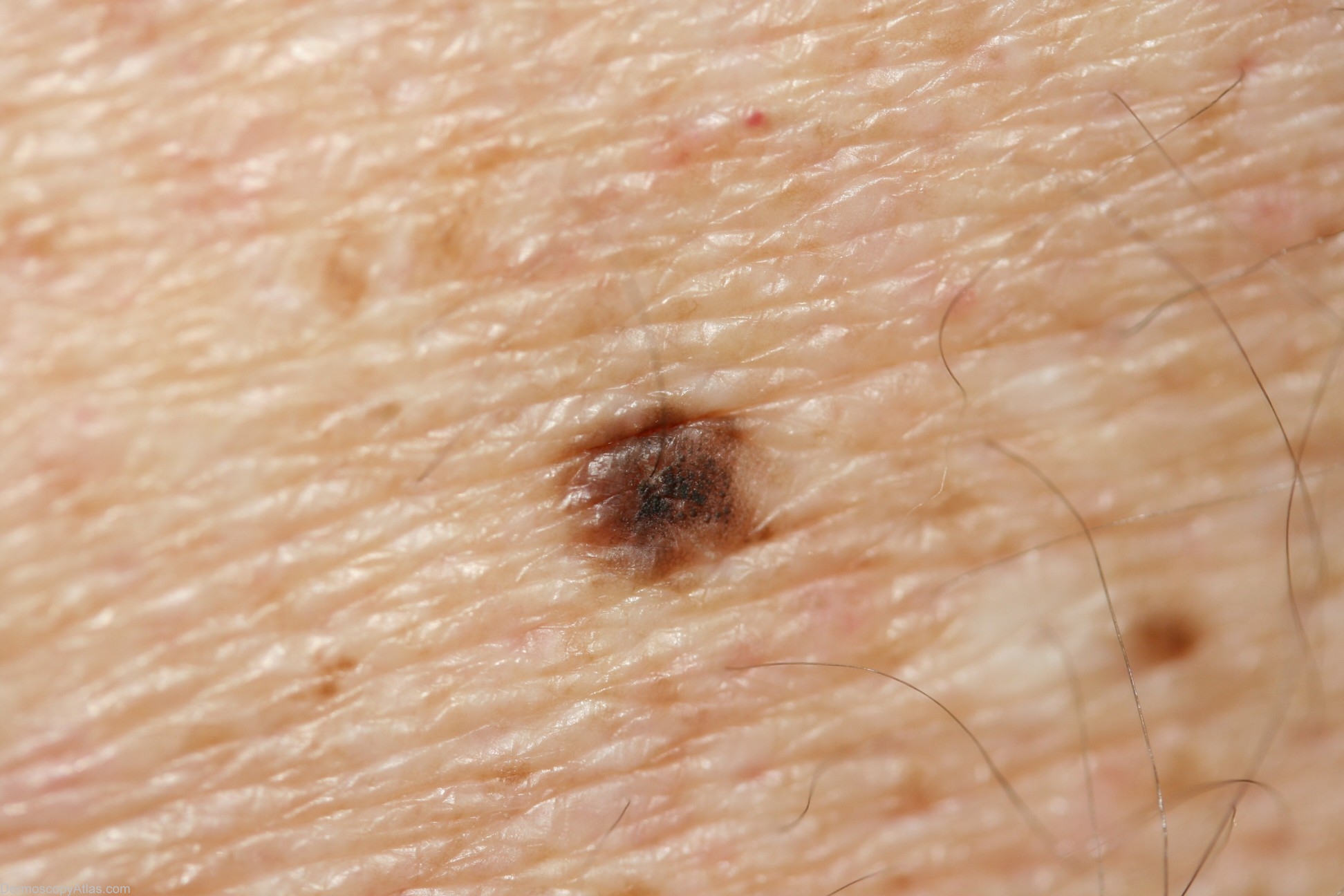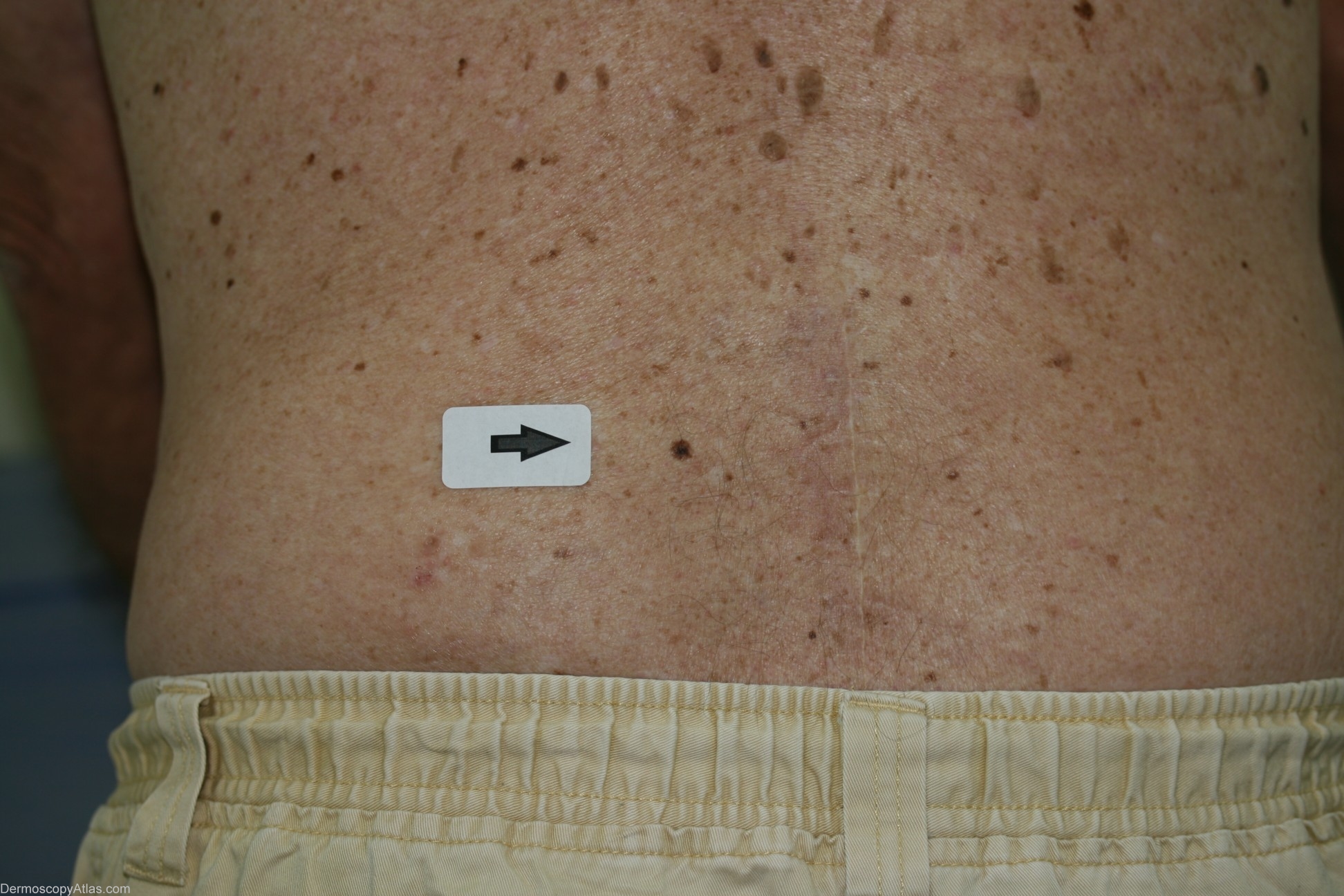
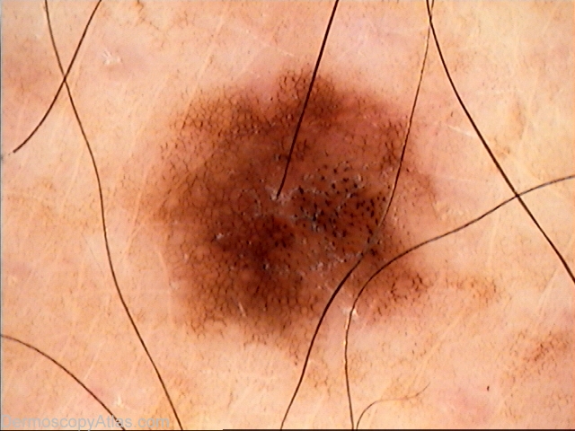
Site: Back
Diagnosis: Nevus dysplastic
Sex: M
Age: 78
Type: Dermlite Non Polarised
Submitted By: Cliff Rosendahl
Description: Original Molemax image
History: This lesion was originally imaged on a Molemax with the intention of monitoring after 3 months. The patient returned after 12 months and because of apparent change the lesion was excised. Histology revealed a naevus with moderate dysplasia and regression, falling short of melanoma in situ.
