Teaching Cases
History
Pattern Analysis continued
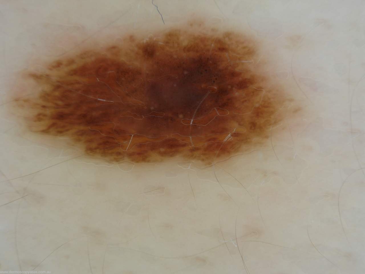
Once you have described the global pattern you look at other structures such as streaks seen at the periphery of lesions or pseudopods which are bulbous projections of the network and are virtually never found in benign lesions.
Peripheral dots of varying colours are more likely in malignant lesions whereas those in the centre of a lesion make it more likely benign.
The blue/white veil should not fill the whole lesion and is frequently found in malignant melanoma over areas of regression.
Regression is shown by both white areas where there is fibrosis and bluish areas where there is melanin and melanophages. Sometimes this is grey in colour. You do not usually get regression in benign lesions. Mind you a lichenoid keratosis is an immune reaction to a solar keratosis or solar lentigo.
Hypo or depigmentation in benign lesions is usually in the centre and is regular whereas it can be anywhere in a melanoma and it is usually irregular.
Blotches are where pigment colour obscures underlying structures. These blotches can be regular or irregular and the more irregular they are the more likely it is a melanoma.
Vascular patterns are important in pattern analysis especially in the diagnosis of amelanotic melanoma and in lesions such as squamous cell skin cancer particularly intraepidermal type.
In melanoma you can get a variety of vascular patterns described as linear irregular and dot vessels. Other vessel patterns include comma, hairpin, arborising and crown shaped.
Pattern analysis can be used to look at all of the skin’s lesions both pigmented and non pigmented and can be used in the diagnosis of basal cell carcinomas, seborrhoeic keratoses and angiomas as well as lesions such as dermatofibromas.
Question:
