Teaching Cases
History
Pattern Analysis
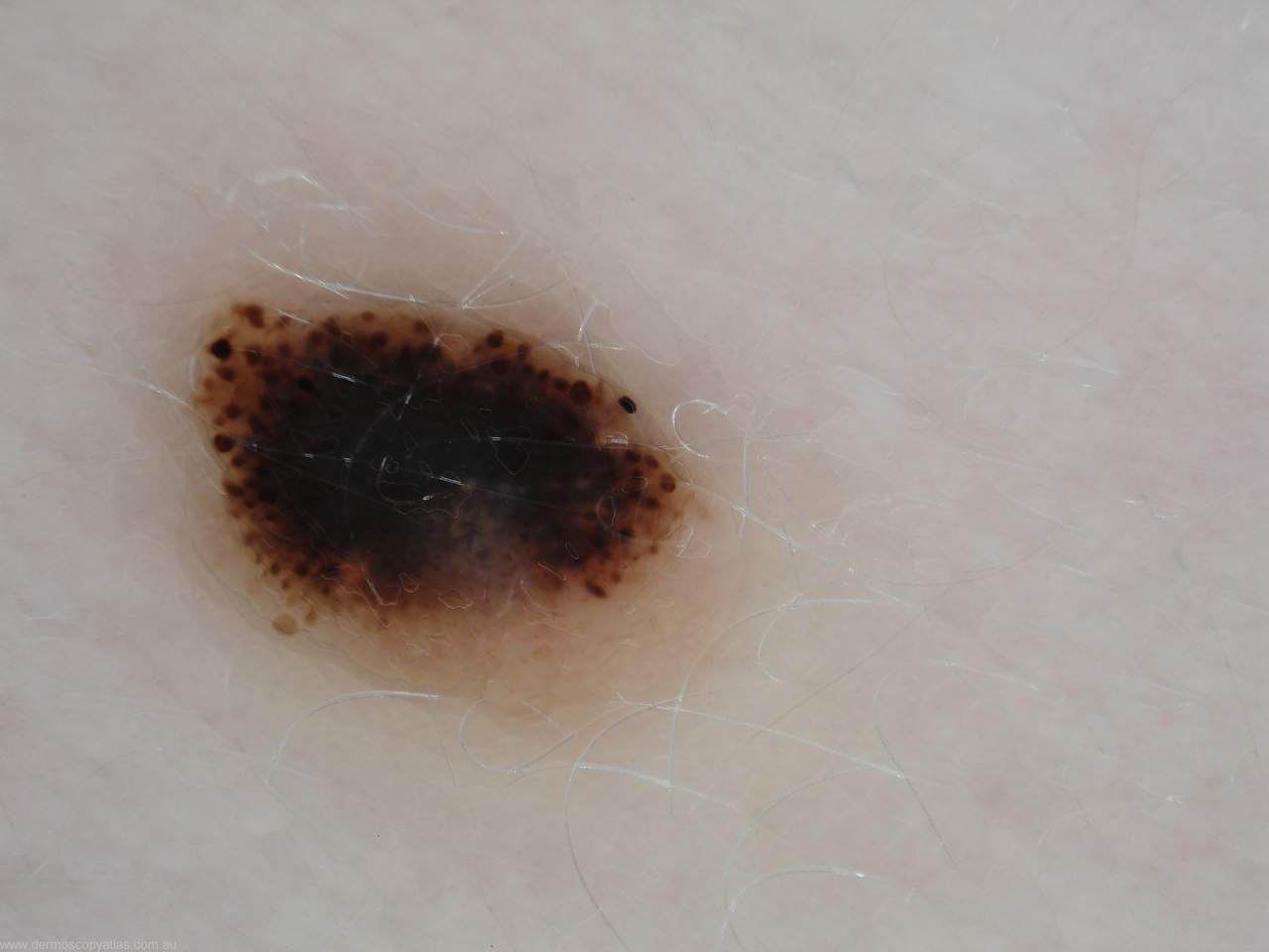
Pattern analysis looks at various components of pigmented lesions including the network, its thickness, whether it has a sharp cut off at the periphery or a gradual cut off, the presence of dots and globules, streaks, blue/white veil, regression, hypopigmentation and also looks carefully at vascular structures. Experienced dermoscopy users are able to look at combinations of these features and determine with great accuracy whether a lesion is benign or malignant.
You first of all look at a pigmented lesion and say whether there is a global pattern. These global patterns include a reticular pattern, described as a netlike structure commonly seen in pigmented lesions, a globular pattern where the globules are uniform in size and regular in benign pigmented lesions but of different sizes and irregularly spaced in dysplastic nevi and melanomas. Globules around the periphery of a lesion are suggestive of an enlarging benign nevus.
A cobblestone pattern refers to angulated globules resembling cobblestones. It is commonly seen in congenital nevi.
Homogeneous pattern has no pigment network or any of these structures and shows just a diffuse uniform colour.
Starburst pattern describes radial streaks from the periphery of the lesion and it is mainly seen in Spitz or Reed nevi.
Parallel pattern is seen on acral lesions on the palms and soles. The parallel ridge pattern commonly signifies a malignant acral melanoma whereas the parallel furrow pattern is seen with benign nevi.
Multi component pattern is a combination of three or more dermoscopic patterns in the same lesion and is supposed to be a feature of melanoma.
Question:
