Teaching Cases
History
Menzies method continued
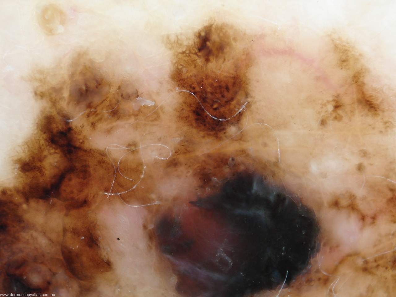
Looking at the positive features radial streaming relates to projections of the edge of a lesion which arises from a network or the tumour itself. Radial streaming is a characteristic of about 18% of melanomas and has a 96% specificity.
Pseudopods are bulbous projections of the edge of a lesion usually arising from the network. They are found in 23% of invasive melanomas and have a specificity of 97%. Their presence is usually very asymmetrical. If they are seen all around the circumference of a lesion it is likely to be a Spitz nevus.
Peripheral black dots and globules represent cellular nests at the proliferating edge of a melanoma. It is the peripheral location of these dots and globules and their dark colour that is highly specific for melanoma. They are found in 42% of invasive melanomas and have a specificity of 92%. The black dots are due to melanin accumulating in the stratum corneum from Pagetoid spread of the melanocytes.
Multiple blue/grey dots represent areas of regression of melanocytic lesions with the melanin being found in melanophages in the dermis. They are found in 45% of invasive melanomas and have a specificity of 91%.
A broadened network refers to an increase in the width of the reticular pigmented net. In melanoma this broadened network is often focal. It is seen in possibly 35% of invasive melanomas with a specificity of 86%. A broadened network is a particular feature of lentigo maligna.
A blue/white veil represents overlying ortho keratosis of the stratum corneum with melanin in the mid dermis. It should not occupy the entire lesion. If it does it is more suggestive of a blue nevus. Blue/white veils are found in 51% of the invasive melanomas and have a specificity of 97%.
Lastly multiple brown dots are seen focally in melanomas. They are found in 30% of melanomas and have a specificity of 97%.
Scar like depigmentation indicates regression within a melanoma. Involved areas are usually pure white in colour and differ from the normal surrounding skin because all the pigment cells have been knocked out. It is seen in 36% of invasive melanomas and has a specificity of 93%. <
B>Multiple colours are certainly a feature of melanoma because of the different levels at which melanin is seen in both the epidermis and the dermis in melanomas. In Menzies system for it to be a significant positive feature there have to be at least five colours from the group comprising red, tan, dark brown, black, grey and blue. They are found in 53% of invasive melanomas and have a specificity of 92%.
Question: Where is Scott Menzies based?
