Teaching Cases
History
The Menzies Method of Dermoscopy
The Menzies method of diagnosing pigmented lesions looks initially to see if the lesion is symmetrical in the nature of the pigment distribution and if there is a single colour. If both of these criteria are present then it is a benign pigmented lesion. If they are not present you then look for certain features that make melanoma more likely. One major and a couple of these minor factors makes the diagnosis of melanoma a likely possibility.
NEGATIVE FEATURES Symmetry of Pigment Pattern
This refers to the pattern of pigmentation and not the shape of the lesion. If the lesion is divided both horizontally and vertically through its long and short axes then there should be symmetry of pigmentation either side of the line. This symmetry is a feature of benign pigmented lesions.
Single Colour
A single colour makes a diagnosis of melanoma unlikely. In melanoma melanocytes are still producing melanin and this melanin is at different levels in the skin ranging from the stratum corneum where it is black to dark brown in the mid epidermis, tan at dermoepidermal junction, grey in the upper dermis and blue in the mid dermis. Colours considered in the Menzies method are black, grey, blue, red, dark brown and tan but white is not scored.
The Menzies method is particularly sensitive in picking melanoma. In other words in using this method you make fewer mistakes when you say something is a melanoma. Symmetry of pigmentation pattern and the presence of only a single colour makes it extremely unlikely that the lesion that you are looking at is a melanoma. These are described as negative features for melanoma. Neither should be found in a melanoma.
POSITIVE FEATURES
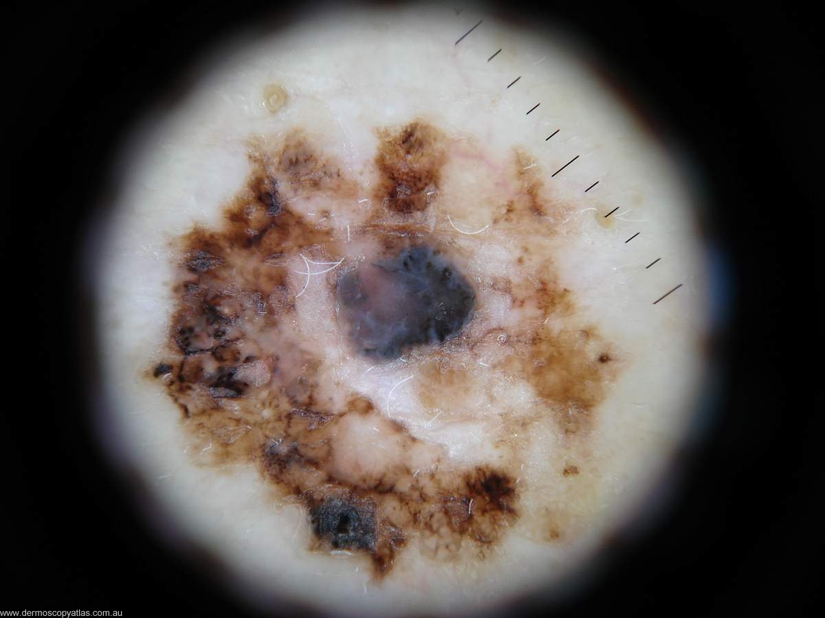
include a blue/white veil, multiple brown dots, radial streaming, pseudopods, scar like depigmentation, peripheral black dots and globules, multiple blue/grey dots or peppering, a broadened network and multiple five to six colours.
To make a diagnosis of melanoma it should have neither of the negative features and at least one of the positive features. The good thing about this method is there is no grading involved. You either have a feature or you don’t. The symmetry of pigment pattern includes both structures and colour along any axis through the centre of the lesion. It does not matter if it is an asymmetrical shape. It is the symmetry of the pigmentation pattern that is important. This is the feature of a benign pigmented lesion. As far as single colours go white is not scored. The presence of a single colour virtually excludes a diagnosis of melanoma. The colour of melanoma varies with the level of the presence of melanin in the epidermis and in the dermis. In the superficial epidermis it is black going through brown to the deep epidermis when it is blue. Melanomas generally have more than one colour.
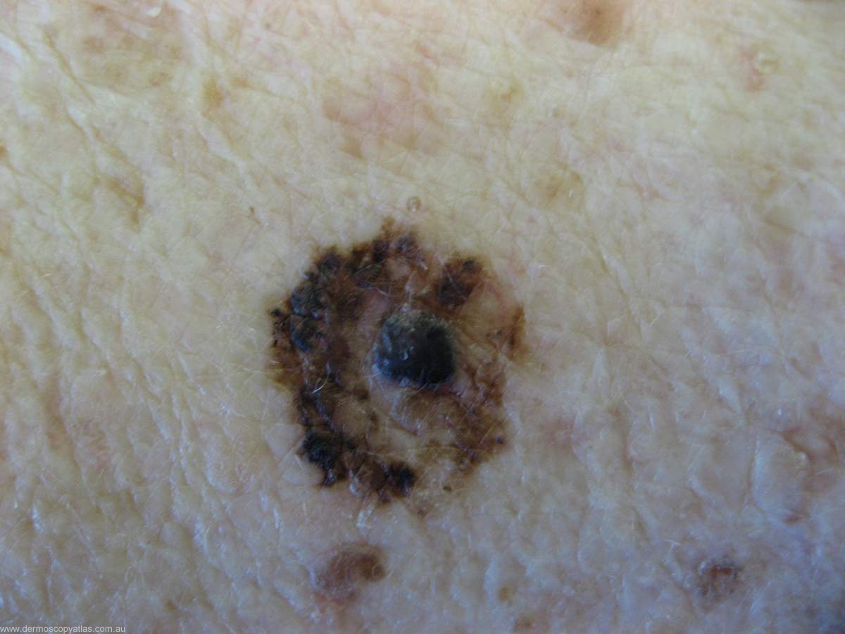
Question: How would you interprit this melanocytic lesion using the Menzies method?

