Loading
TeleDermConsult-logo
  • Recent Submissions
  • Search
  • Diagnosis
  • Quiz
  • Image comparison
  • Submit
  • Teaching Cases
  • Index
  • About

Teaching Cases


teach1

History

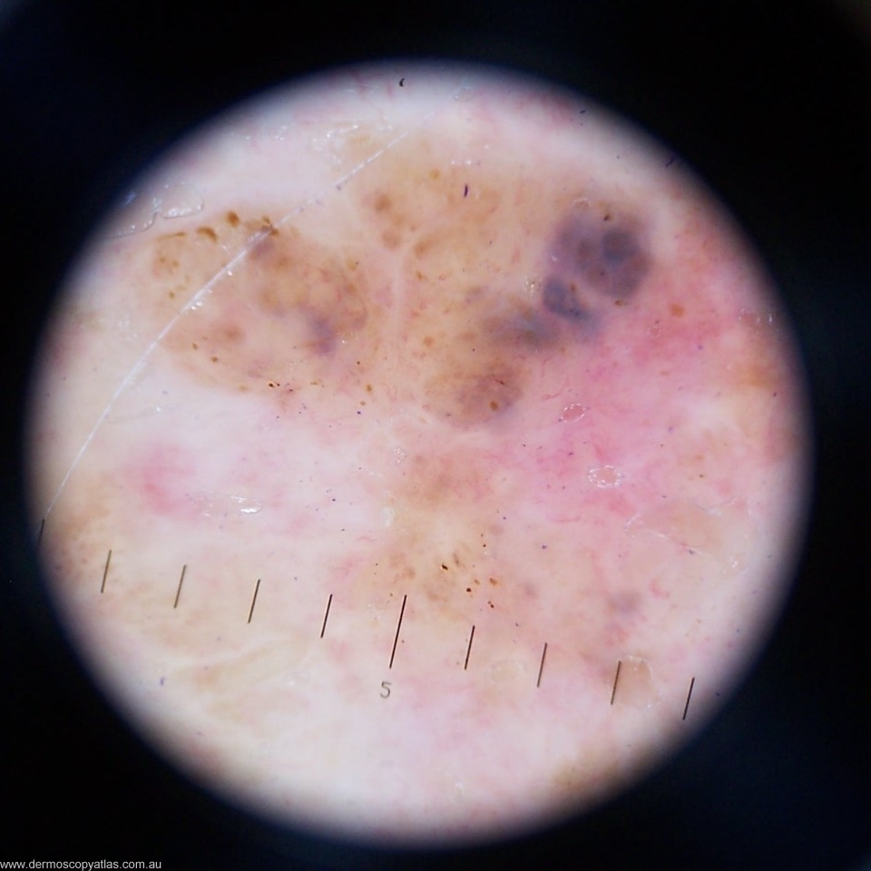
Case 3. This 79 year old patient with type 1 skin has had > 100 NMSC. This apparently new lesion on the anterior shoulder was noted at a 6 monthly check. Consider Solar lentigo, Seborrhoeic keratosis, Pigmented BCC, Nevus, Melanoma or Other.

teach1

View Full Size Image

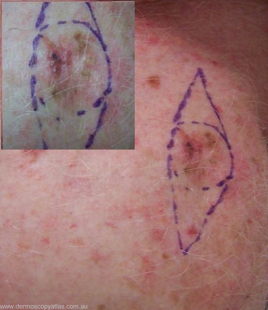
Question: What is your clinical diagnosis?

Answer: Clinically this looks suspicious. It is a pigmented lesion with varying colours and a suggestion of regression.


Question: What is your dermatoscopic diagnosis?

Answer: There is no obvious network but the brown dots make it melanocytic. The amorphous blue area is suggestive of BCC but the adjacent pink area would be more likely in melanoma. The linear irregular vessels more support melanoma.


Question: What do you think the histology will show?

Answer: Histology showed " Sections show a superficial spreading malignant melanoma. The lesion is mostly in situ but does include a component which is Level 3 and 0.9 mm thick. Rare dermal mitotic figures are seen (less than 3 per square mm). No ulceration, regr


Next Question
  • Recent Submissions
  • Search
  • Diagnosis
  • Quiz
  • Image comparison
  • Submit an Image
  • Teaching Cases
  • Index
  • About

Copyright 2022. Dermoscopy Atlas. ALL Rights Reserved.

Change Image