History
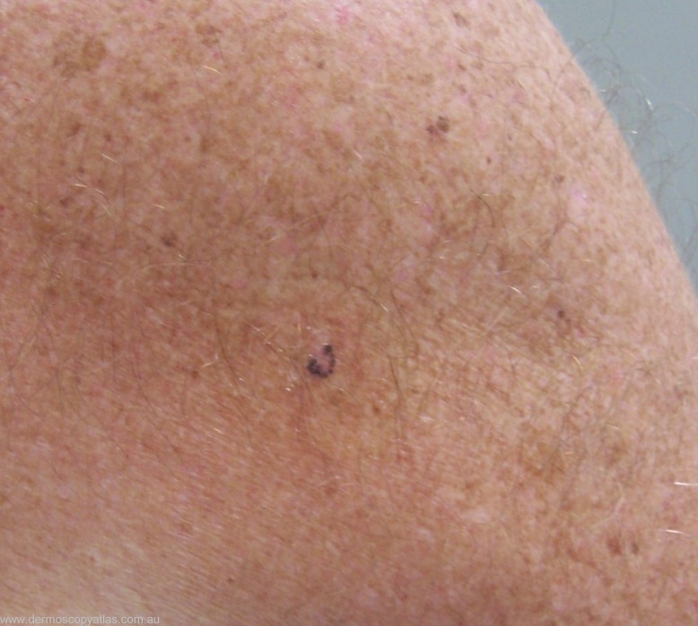
This odd shaped lesion was noted during an rountine physical check up. The patient is 69 years old and has no history of skin cancer. What is your clinical diagnosis?
Consider Melanoma, BCC, IEC and Foreign body reaction as well as Other!
Question: What is your clinical diagnosis?
Answer: Clinically this is a heavily pigmented lesion with a pink background. You would consider pigmented BCC or Melanoma.
Question: What is your dermatoscopic diagnosis?
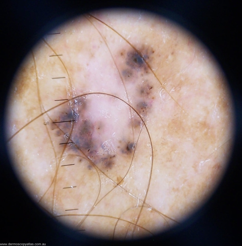
Answer: There is no network but the brown dots suggest this is melanocytic and the white area might be regression. The blue amorphous areas however more suggest pigmented BCC.
Question: What do you think the histology will show?
Answer: The Histology was reported as follows-Histology showed superficial multifocal BCC, with no comment regarding pigmentation. Surely some comment about pigmentation would have been appropriate!


