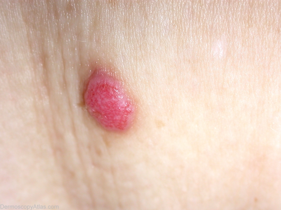
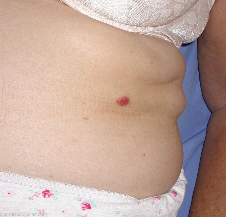
Loading
TeleDermConsult-logo
  • Recent Submissions
  • Search
  • Diagnosis
  • Quiz
  • Image comparison
  • Submit
  • Teaching Cases
  • Index
  • About

<< Back to Diagnosis Page

Image Number #2179 (Clear cell acanthoma)

Site: Chest

Diagnosis: Clear cell acanthoma

Sex: M

Age: 56

Type: Heine

Submitted By: Ian McColl

View Full Size Image

Click Here to View Details

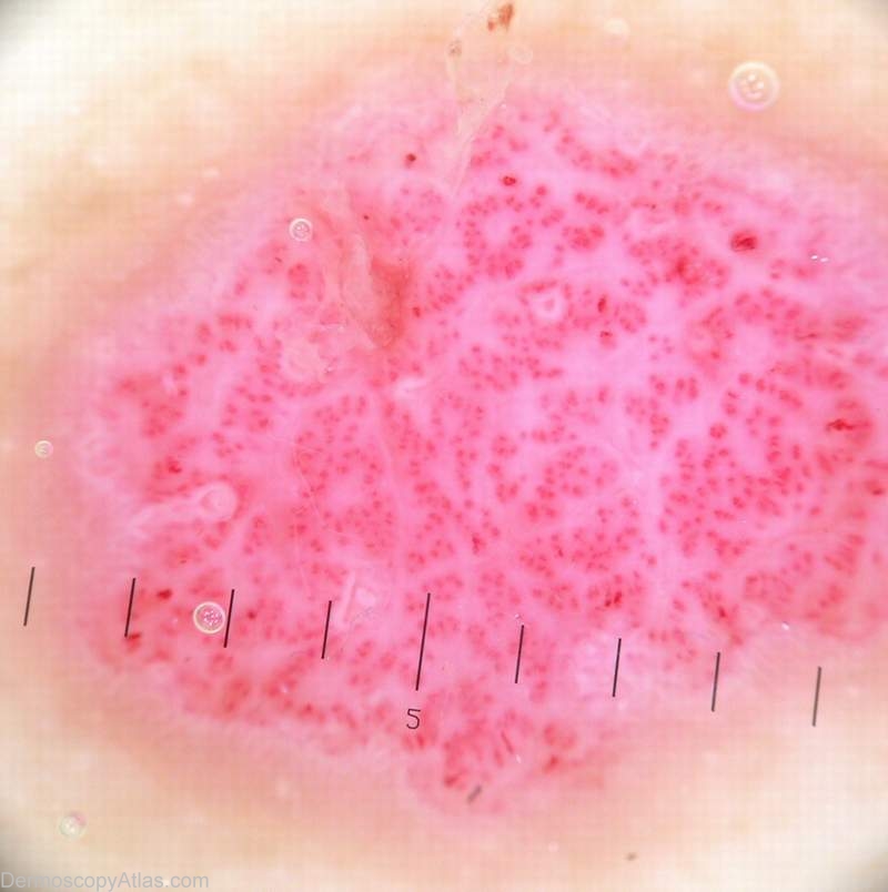
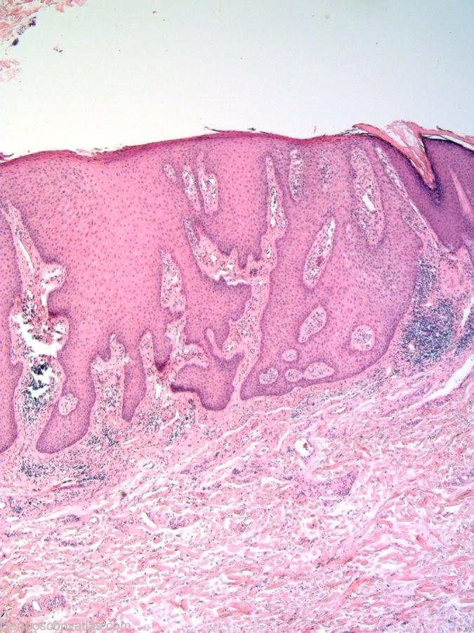
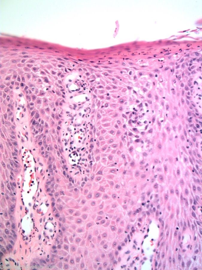
Description: Histopathology. The pale keratinocytes will take up a PAS stain showing glycogen in the cells.Pale cells in an acanthotic epidermis can also be seen in clonal seborrhoeic keratoses and some warts. Other epidermal changes include mild spongiosis, exocytosis of neutrophils into the epidermis which may rarely form intraepidermal microabscesses.

History: Images courtesy of Dr Alan Cameron This lesion arose slowly over a 16 week period. There was no bleeding.
View the Blog discussion of this case

DermNetNZ   eMedicine   PubMed   Dermatology Online   Archives   JAAD for "Clear cell acanthoma"

  • Recent Submissions
  • Search
  • Diagnosis
  • Quiz
  • Image comparison
  • Submit an Image
  • Teaching Cases
  • Index
  • About

Copyright 2022. Dermoscopy Atlas. ALL Rights Reserved.

Change Image