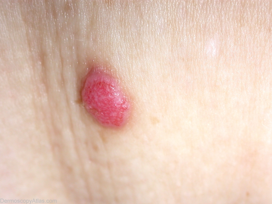
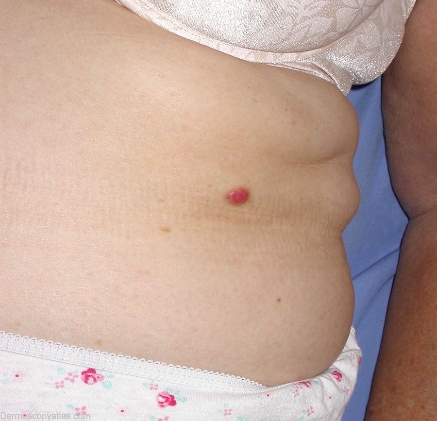
Loading
TeleDermConsult-logo
  • Recent Submissions
  • Search
  • Diagnosis
  • Quiz
  • Image comparison
  • Submit
  • Teaching Cases
  • Index
  • About

<< Back to Diagnosis Page

Image Number #2178 (Clear cell acanthoma)

Site: Chest

Diagnosis: Clear cell acanthoma

Sex: M

Age: 56

Type: Heine

Submitted By: Ian McColl

View Full Size Image

Click Here to View Details

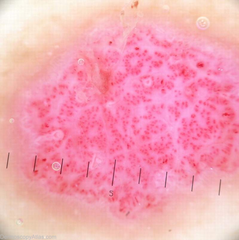
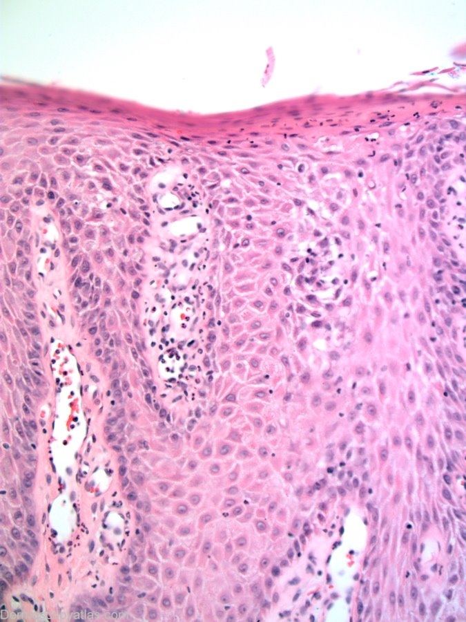
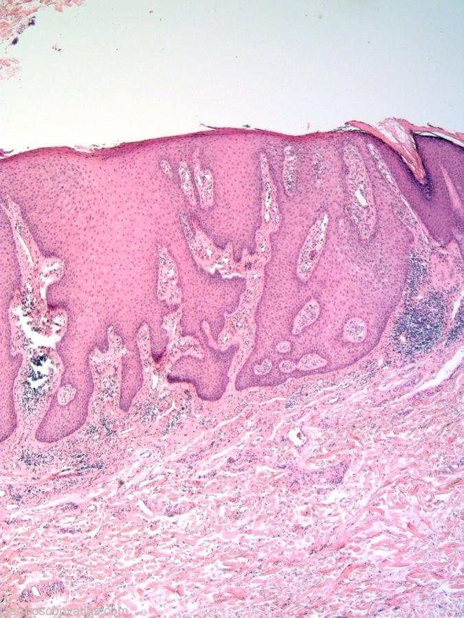
Description: Histopathology. This lesion is considered as a benign epidermal neoplasm.There is psoriasiform hyperplasia with pale staining keratinocytes.The acanthotic downgrowths often fuse. There is a mixed inflammatory cell infiltrate.

History: Images courtesy of Dr Alan Cameron This lesion arose slowly over a 16 week period. There was no bleeding.
View the Blog discussion of this case

DermNetNZ   eMedicine   PubMed   Dermatology Online   Archives   JAAD for "Clear cell acanthoma"

  • Recent Submissions
  • Search
  • Diagnosis
  • Quiz
  • Image comparison
  • Submit an Image
  • Teaching Cases
  • Index
  • About

Copyright 2022. Dermoscopy Atlas. ALL Rights Reserved.

Change Image