Loading
TeleDermConsult-logo
  • Recent Submissions
  • Search
  • Diagnosis
  • Quiz
  • Image comparison
  • Submit
  • Teaching Cases
  • Index
  • About

<< Back to Diagnosis Page

Image Number #3580 (Lentigo Maligna)

Site: Cheek

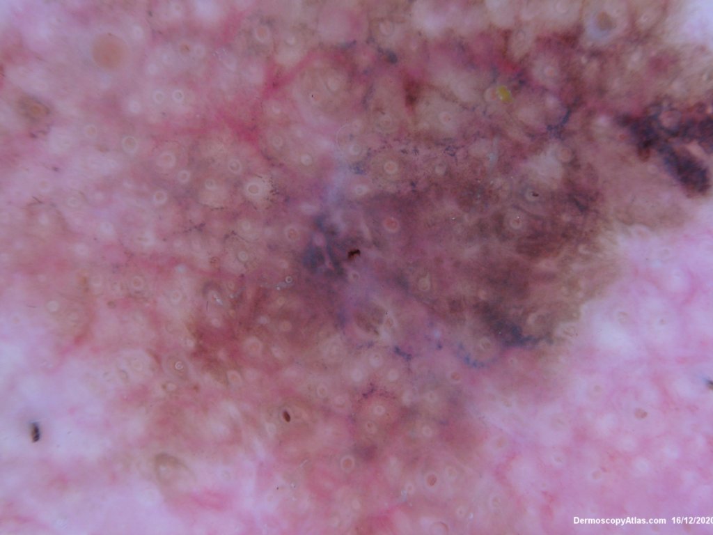
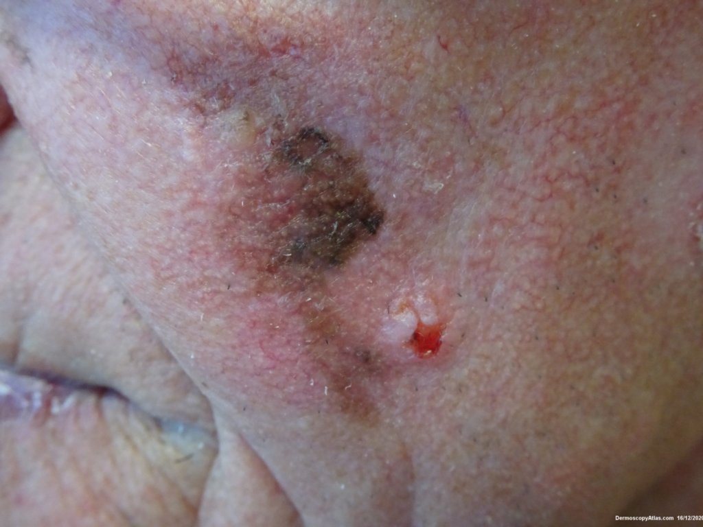
Diagnosis: Lentigo Maligna

Sex: M

Age: 80

Type: Dermlite Polarised

Submitted By: Ian McColl

View Full Size Image

Click Here to View Details

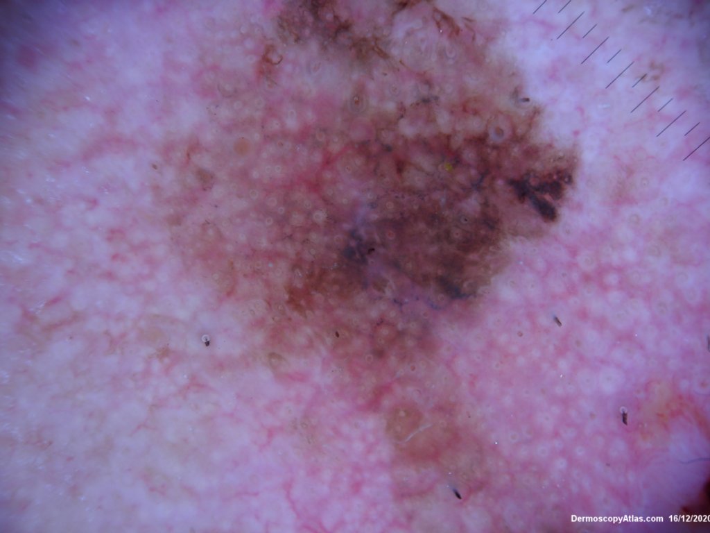
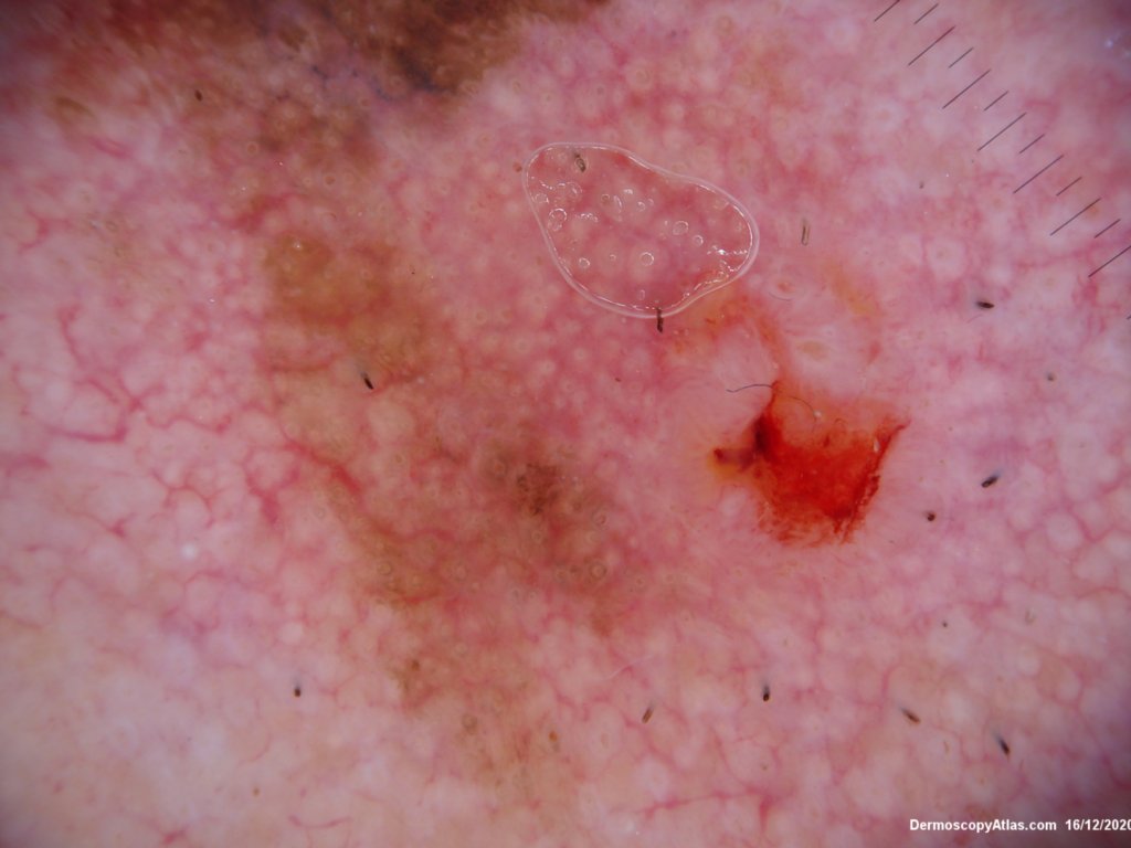
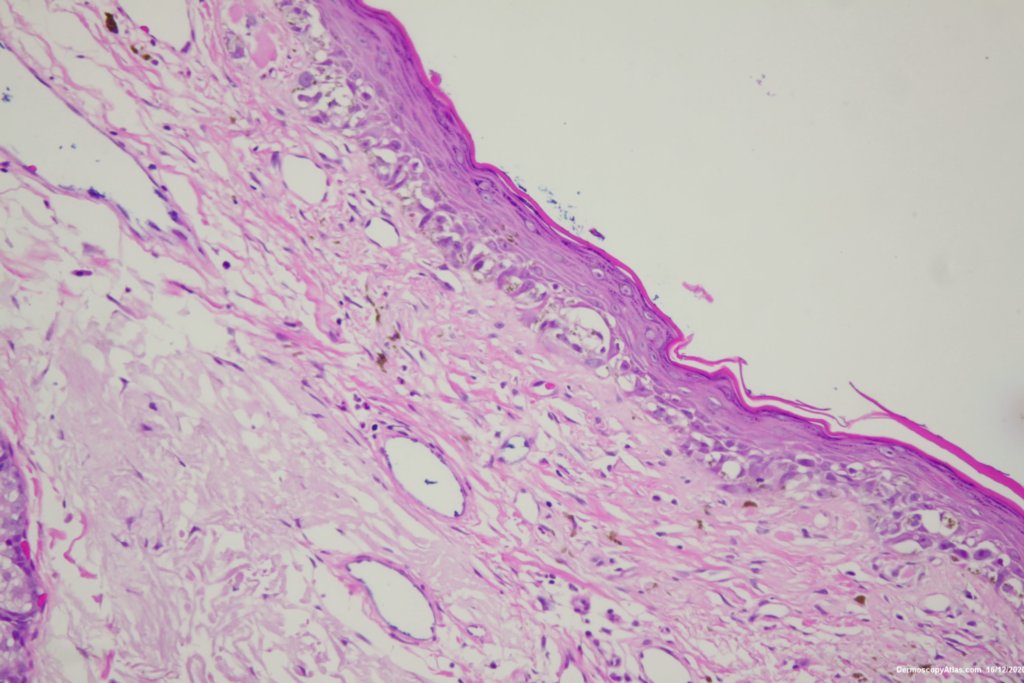
Description: Grey circles, grey dots, perifollicular pigmentation

History:

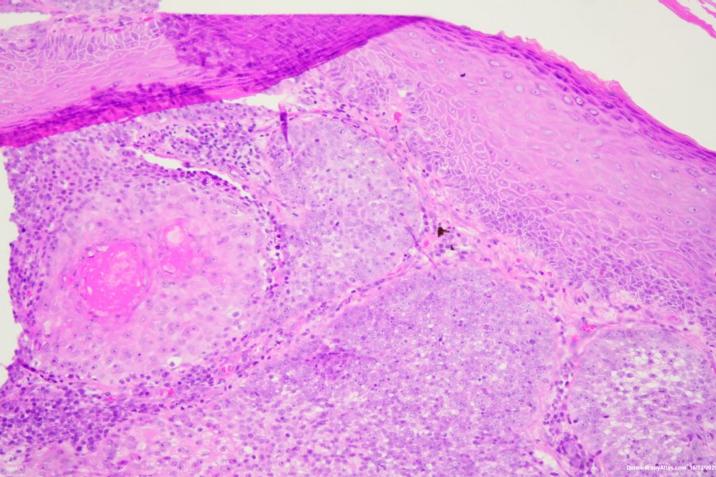
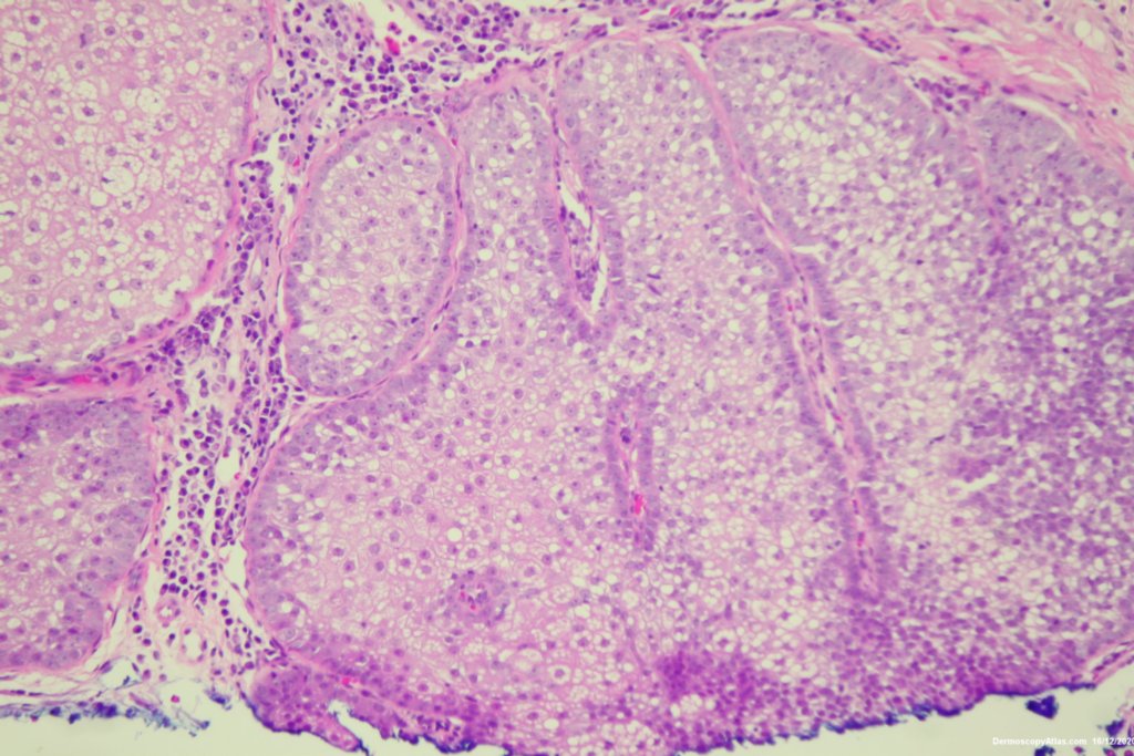
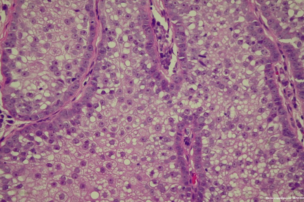
 Elderly male with no past history of melanoma developed these two lesions over a year or so. The lentigo maligna was easy to diagnose on the clinical and dermatoscopy but the nodule was more difficult. Was it an amelanotic melanoma, BCC or SCC were the initial thoughts. Histology showed a well differentiated sebaceous carcinoma. The two lesions were excised in the same specimen with 5 mm margins. Studies failed to show the mutation for the Muir Torre syndrome which can be seen with sebaceous carcinoma.

See Lentigo maligna in Dermoscopy Made Simple

EyeWiki on Sebaceous Carcinoma

DermNetNZ   eMedicine   PubMed   Dermatology Online   Archives   JAAD for "Lentigo Maligna"

  • Recent Submissions
  • Search
  • Diagnosis
  • Quiz
  • Image comparison
  • Submit an Image
  • Teaching Cases
  • Index
  • About

Copyright 2022. Dermoscopy Atlas. ALL Rights Reserved.

Change Image