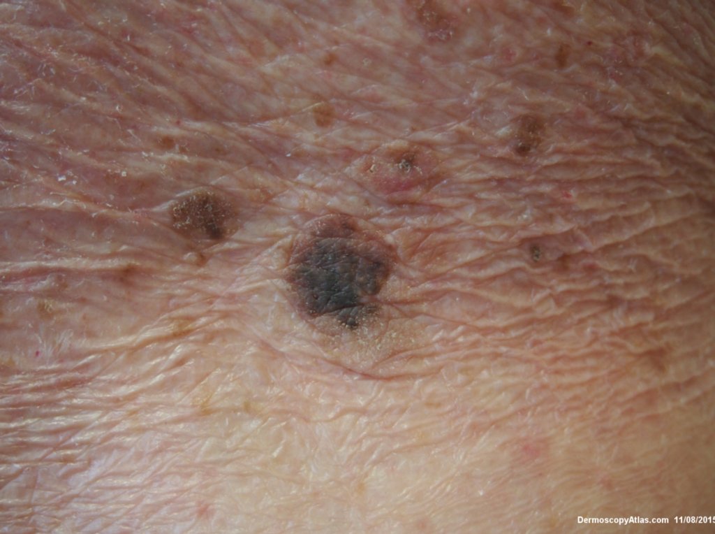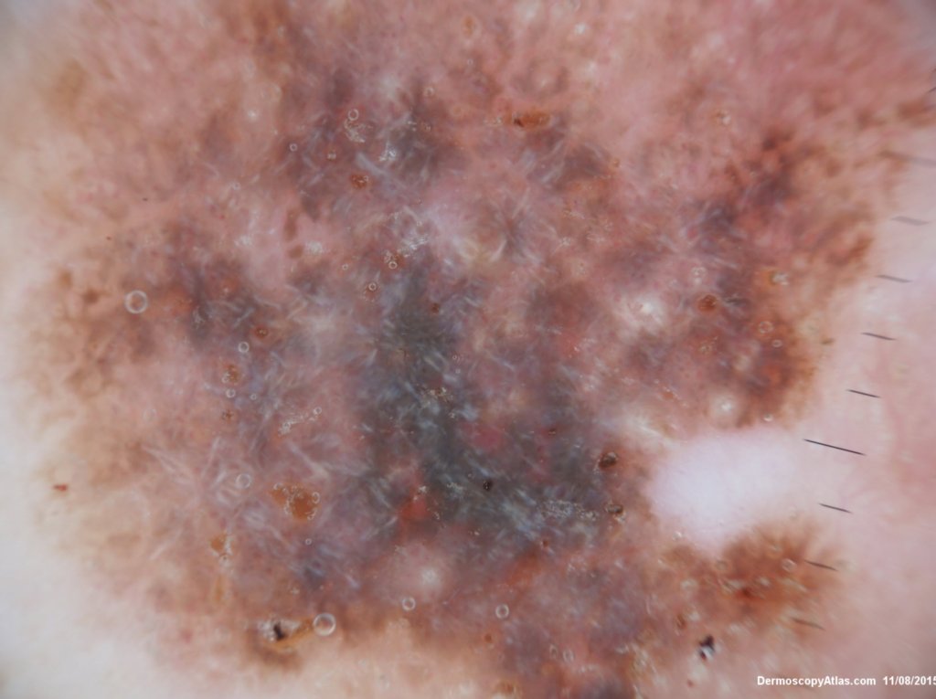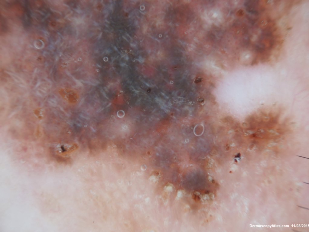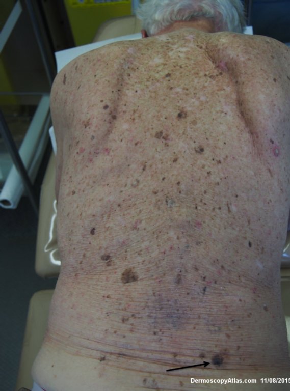
Site: Back
Diagnosis: Seborrhoeic keratosis irritated
Sex: M
Age: 92
Type: Dermlite Polarised
Submitted By: Ian McColl
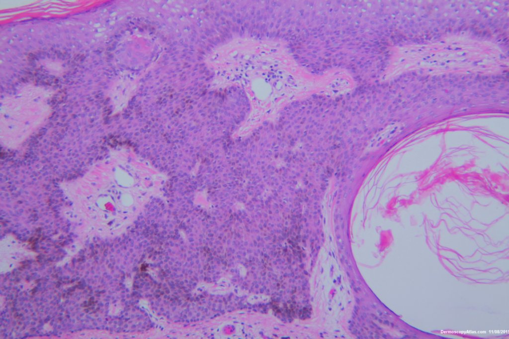
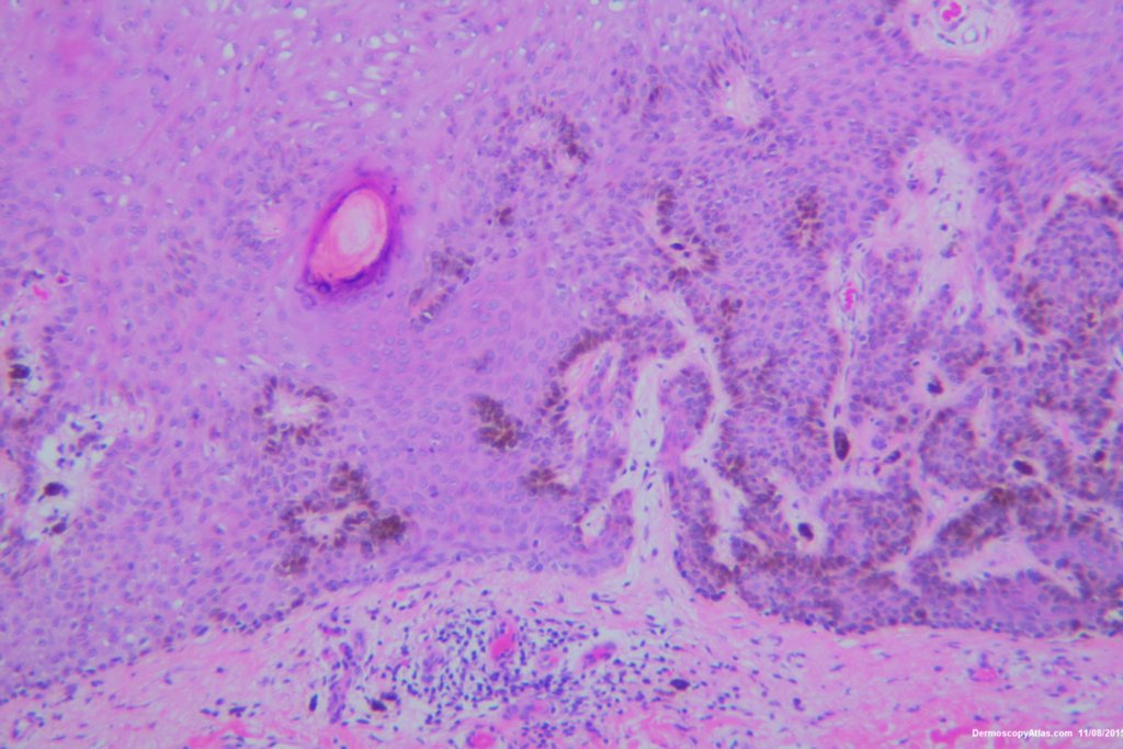
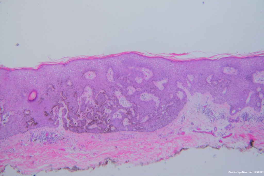
Description: Pathology of Pigmented seborrhoeic keratosis
History:
This lesion looked darker than the many other seborrhoeic keratoses on this elderly man's back. It was thought there might be a lentiginous proliferation of atypical melanocytes in the lesion . Also the multiple colours and the white lines were unusual for a seborrhoeic keratosis. However pathology of a large shave biopsy excision showed only a pigmented thick seborrhoeic keratosis with increased pigment in normal basal keratinocytes. The blue colour was because of the depth of the lesion ie thick. No idea why the white lines!
