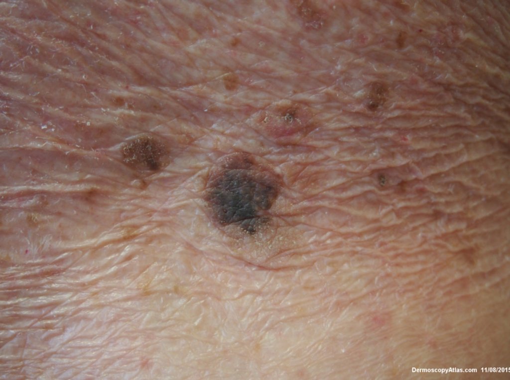
Loading
TeleDermConsult-logo
  • Recent Submissions
  • Search
  • Diagnosis
  • Quiz
  • Image comparison
  • Submit
  • Teaching Cases
  • Index
  • About

<< Back to Diagnosis Page

Image Number #3374 (Seborrhoeic keratosis irritated)

Site: Back

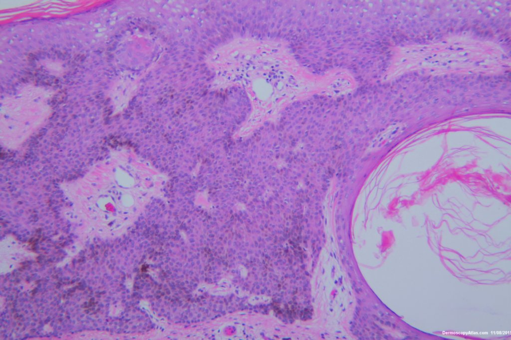
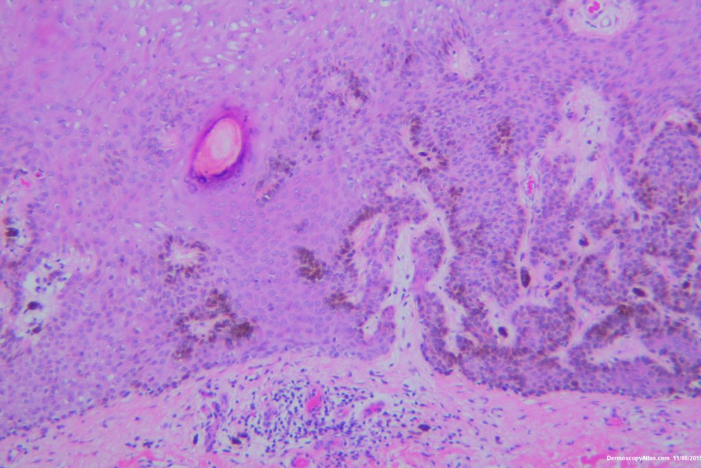
Diagnosis: Seborrhoeic keratosis irritated

Sex: M

Age: 92

Type: Dermlite Polarised

Submitted By: Ian McColl

View Full Size Image

Click Here to View Details

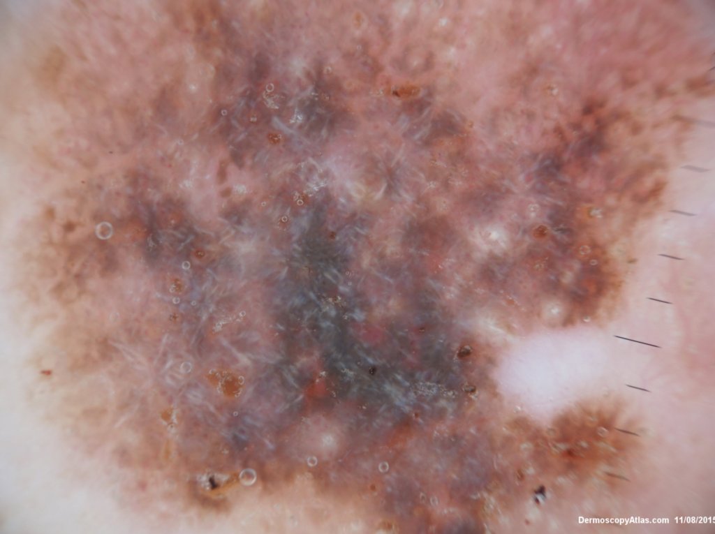
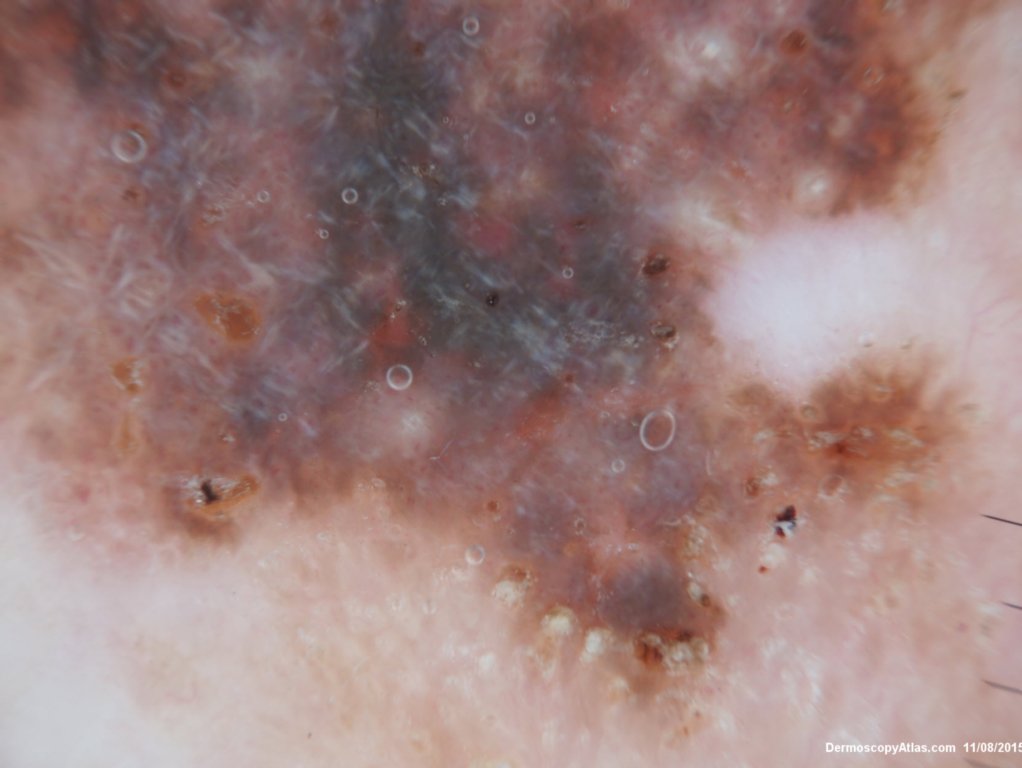
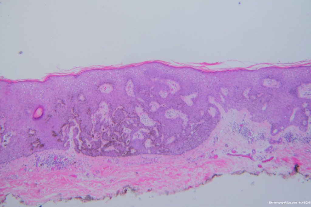
Description: Pigmented lesion back

History:

 This lesion looked darker than the many other seborrhoeic keratoses on this elderly man's back. It was thought there might be a lentiginous proliferation of atypical melanocytes in the lesion . Also the multiple colours and the white lines were unusual for a seborrhoeic keratosis. However pathology of a large shave biopsy excision showed only a pigmented thick seborrhoeic keratosis with increased pigment in normal basal keratinocytes. The blue colour was because of the depth of the lesion ie thick. No idea why the white lines!

View the video of the pathology here.

DermNetNZ   eMedicine   PubMed   Dermatology Online   Archives   JAAD for "Seborrhoeic keratosis irritated"

  • Recent Submissions
  • Search
  • Diagnosis
  • Quiz
  • Image comparison
  • Submit an Image
  • Teaching Cases
  • Index
  • About

Copyright 2022. Dermoscopy Atlas. ALL Rights Reserved.

Change Image