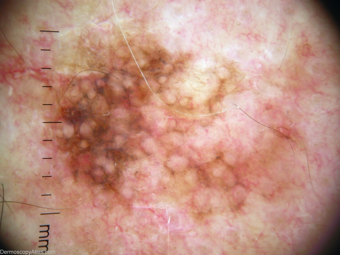
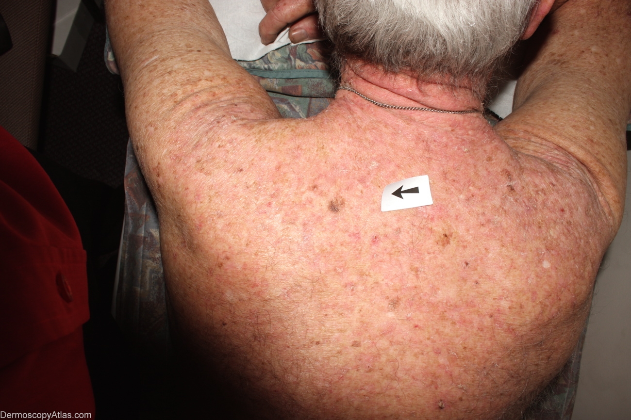
Loading
TeleDermConsult-logo
  • Recent Submissions
  • Search
  • Diagnosis
  • Quiz
  • Image comparison
  • Submit
  • Teaching Cases
  • Index
  • About

<< Back to Diagnosis Page

Image Number #2962 (Melanoma in situ)

Site: Back

Diagnosis: Melanoma in situ

Sex: M

Age: 53

Type: Dermlite Non Polarised

Submitted By: Alan Cameron

View Full Size Image

Click Here to View Details

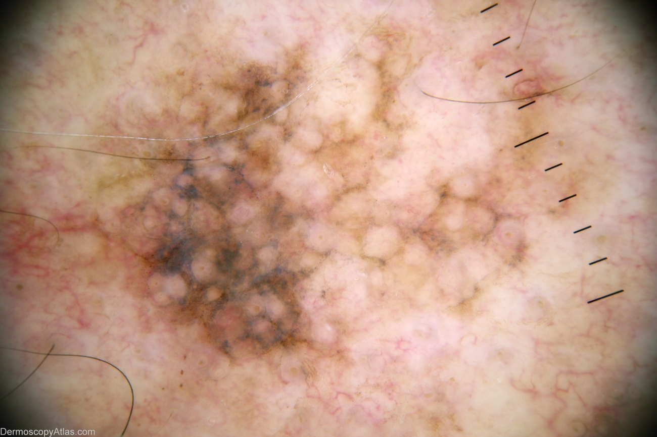
Description: Alternative view, contact polarising dermoscopy

History: 53 year old man with past history multiple BCCs. This lesion noted by GP. Histology reported as; Sections show a level 1 (in situ) lentigo maligna melanoma. There is no ulceratiopn, significant lymphocytic infiltrate or dermal invasion. There is some old regression with melanin inconctinence but no active regression.

DermNetNZ   eMedicine   PubMed   Dermatology Online   Archives   JAAD for "Melanoma in situ"

  • Recent Submissions
  • Search
  • Diagnosis
  • Quiz
  • Image comparison
  • Submit an Image
  • Teaching Cases
  • Index
  • About

Copyright 2022. Dermoscopy Atlas. ALL Rights Reserved.

Change Image