
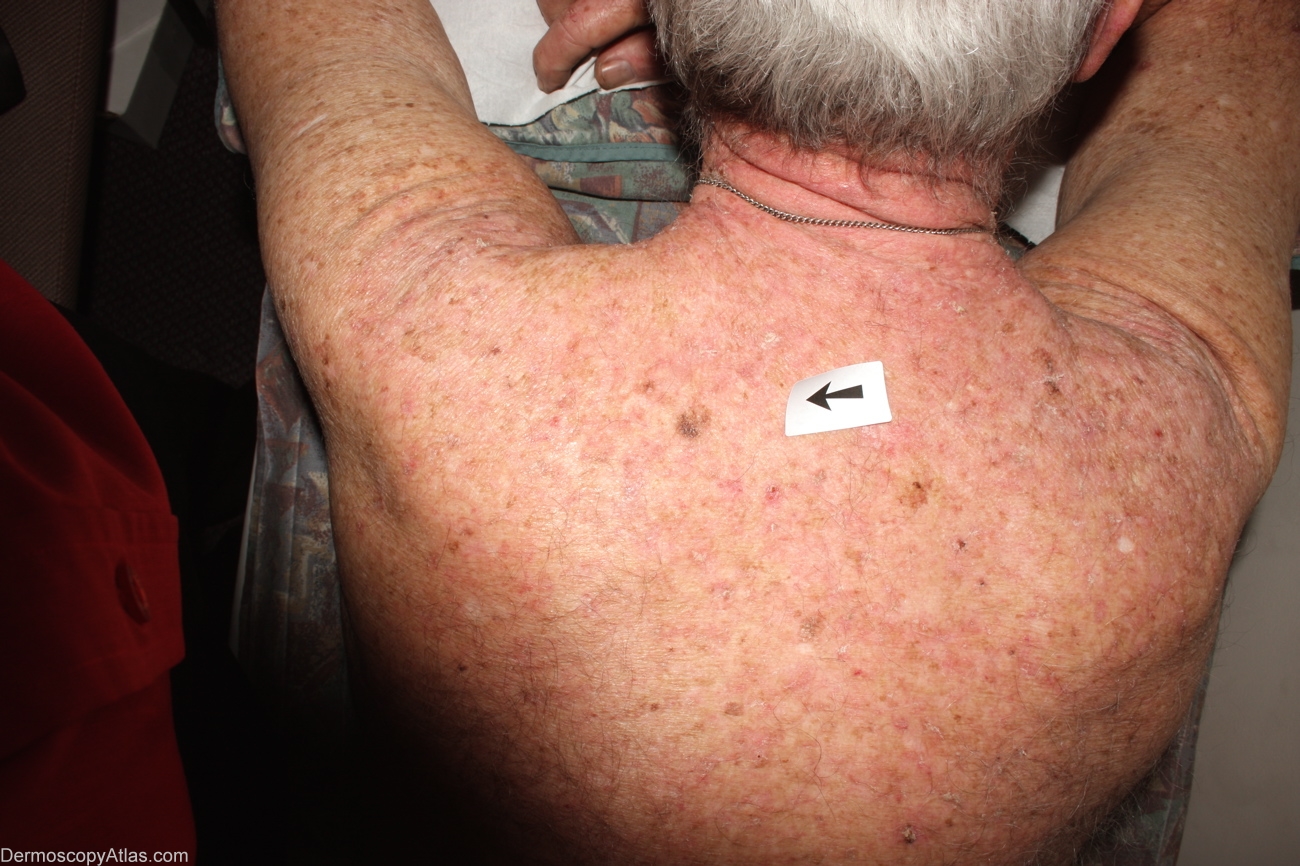
Site: Back
Diagnosis: Melanoma in situ
Sex: M
Age: 53
Type: Dermlite Non Polarised
Submitted By: Alan Cameron
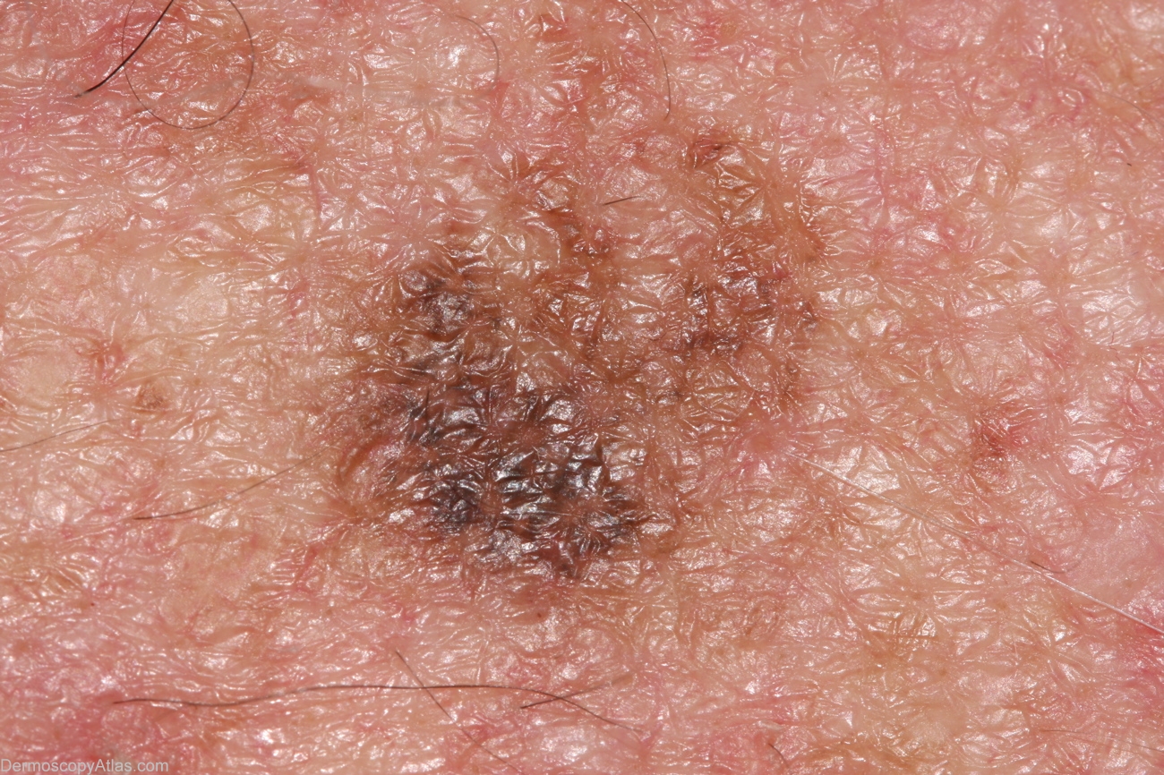
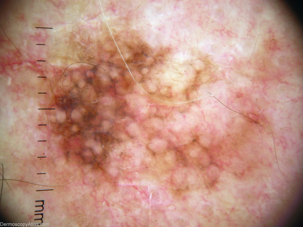
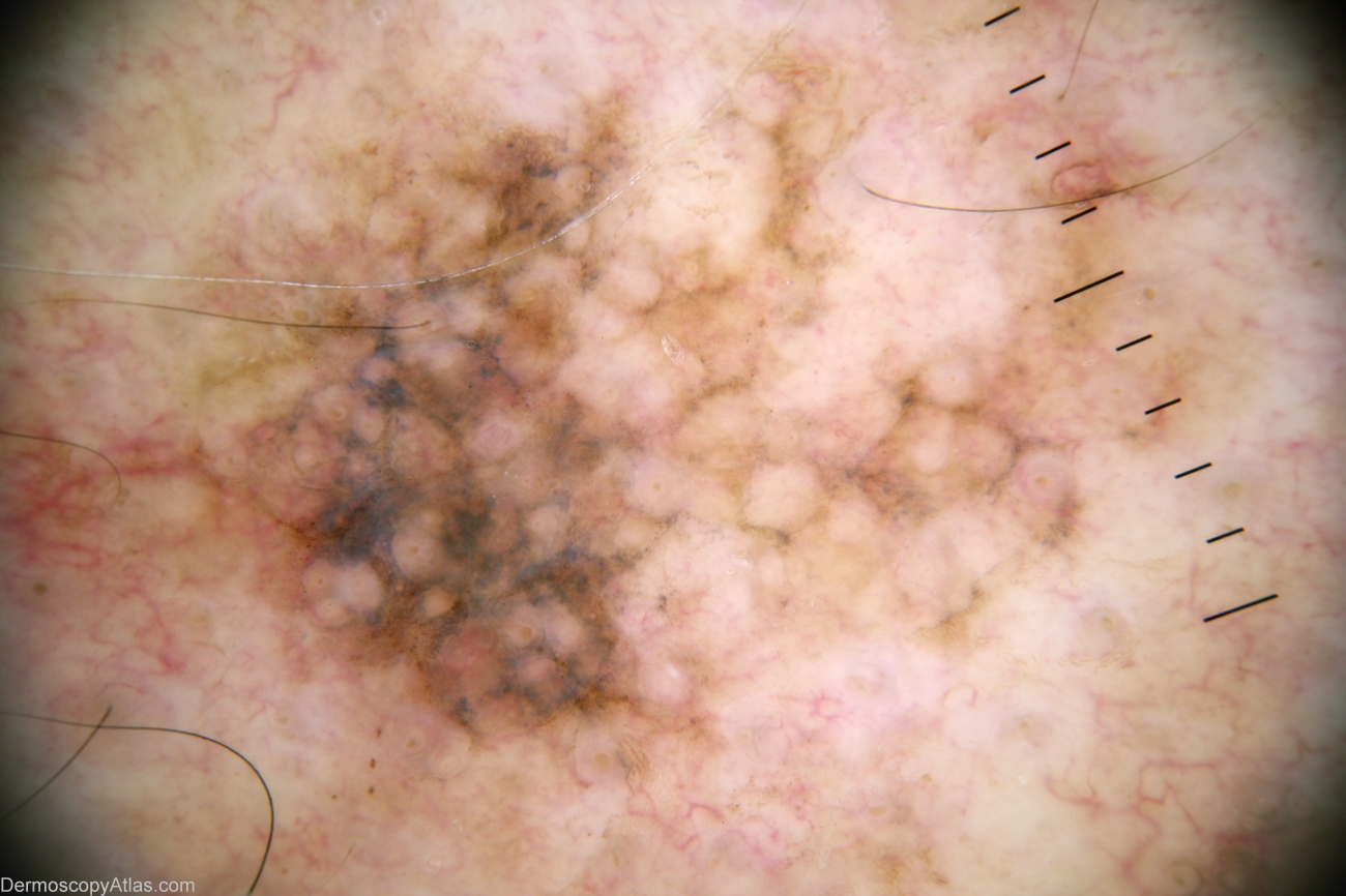
Description: Pattern shows very coarse lines reticular, pink structureless area top right, and grey in a pattern not unlike annular/granular in facial lesions. It has been suggested that heavy actinic damage may efface the rete ridges, giving rise to pseudonetwork, not network. I could not dermoscopically diffentiate between regressing solar lentigo and lentigo maligna.
History: 53 year old man with past history multiple BCCs. This lesion noted by GP. Histology reported as; Sections show a level 1 (in situ) lentigo maligna melanoma. There is no ulceratiopn, significant lymphocytic infiltrate or dermal invasion. There is some old regression with melanin inconctinence but no active regression.