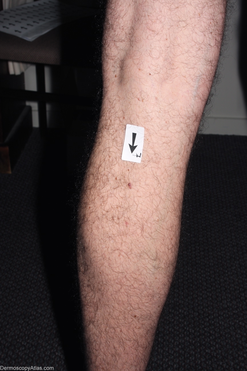
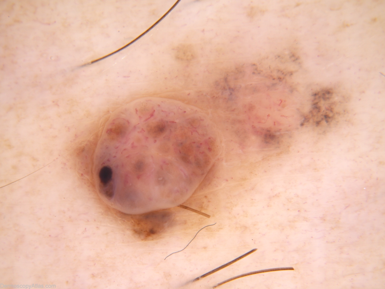
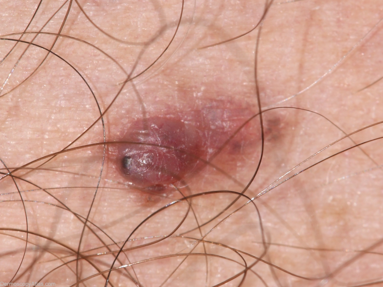
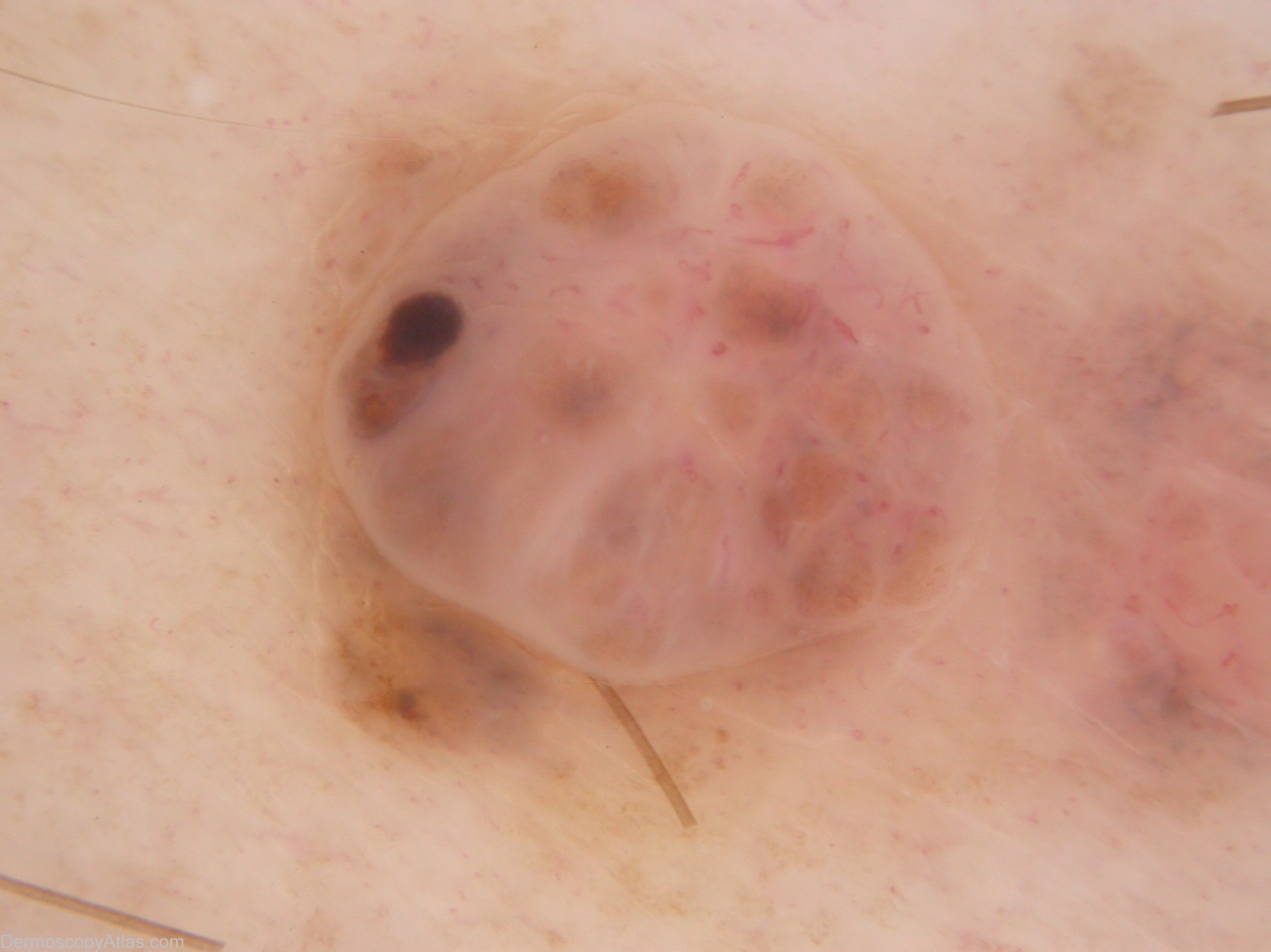
Loading
TeleDermConsult-logo
  • Recent Submissions
  • Search
  • Diagnosis
  • Quiz
  • Image comparison
  • Submit
  • Teaching Cases
  • Index
  • About

<< Back to Diagnosis Page

Image Number #2953 (Melanoma invasive)

Site: Calf

Diagnosis: Melanoma invasive

Sex: M

Age: 35

Type: Dermlite Non Polarised

Submitted By: Alan Cameron

View Full Size Image

Click Here to View Details

Description:

History: 35 year old male with atypical mole syndrome. This lesion noted at examination prompted by melanoma 393. On asking, the patient mentioned this lesion had been irritated by clothing when active for the last 6-9 months. Histology reported as: Sections show a level 3 (0.8mm thick) nodular melanoma with prominent old regression. There is no ulceration, dermal mitoses or lymphocytic infiltrate.

DermNetNZ   eMedicine   PubMed   Dermatology Online   Archives   JAAD for "Melanoma invasive"

  • Recent Submissions
  • Search
  • Diagnosis
  • Quiz
  • Image comparison
  • Submit an Image
  • Teaching Cases
  • Index
  • About

Copyright 2022. Dermoscopy Atlas. ALL Rights Reserved.

Change Image