
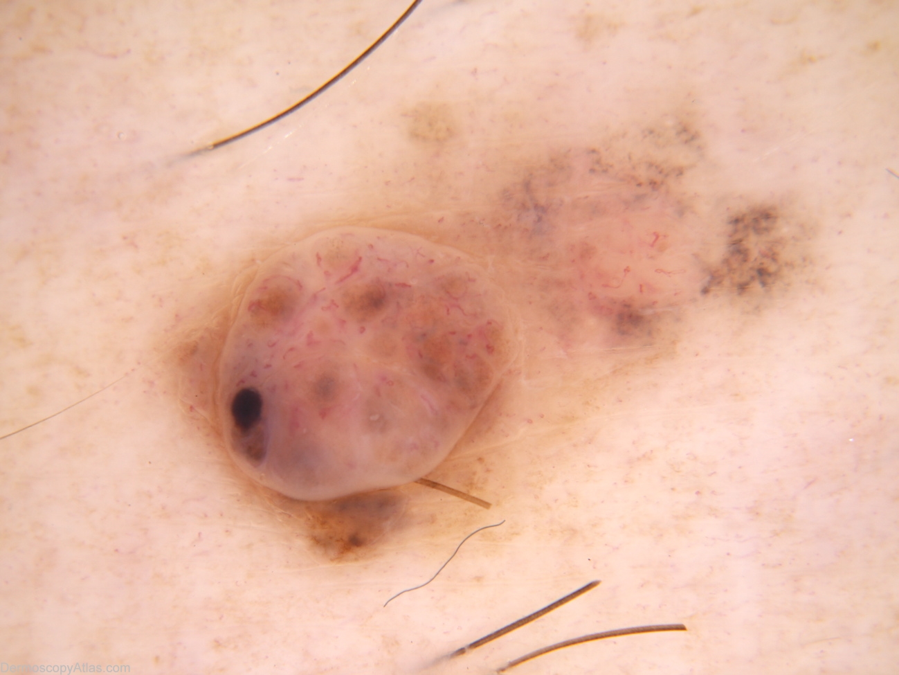
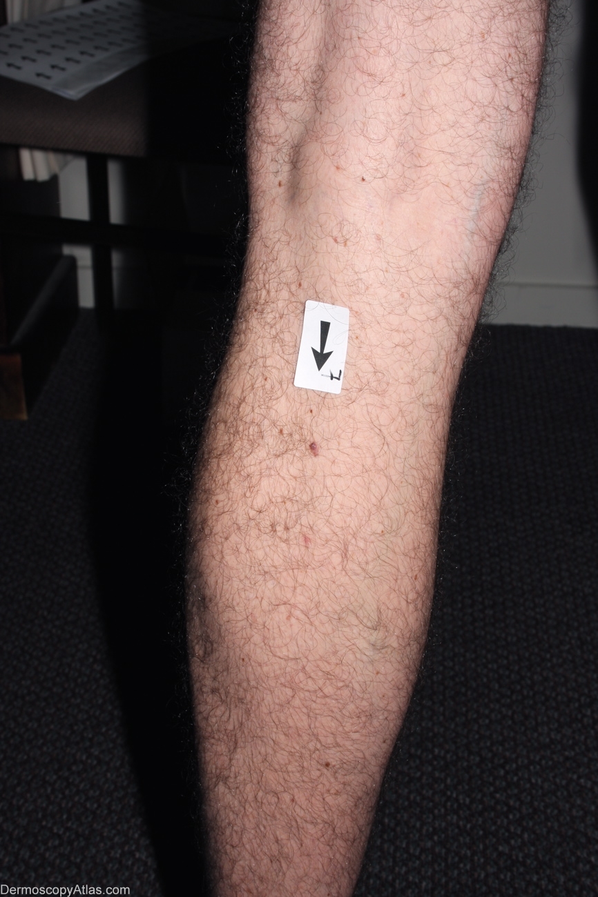
Site: Calf
Diagnosis: Melanoma invasive
Sex: M
Age: 35
Type: Dermlite Non Polarised
Submitted By: Alan Cameron
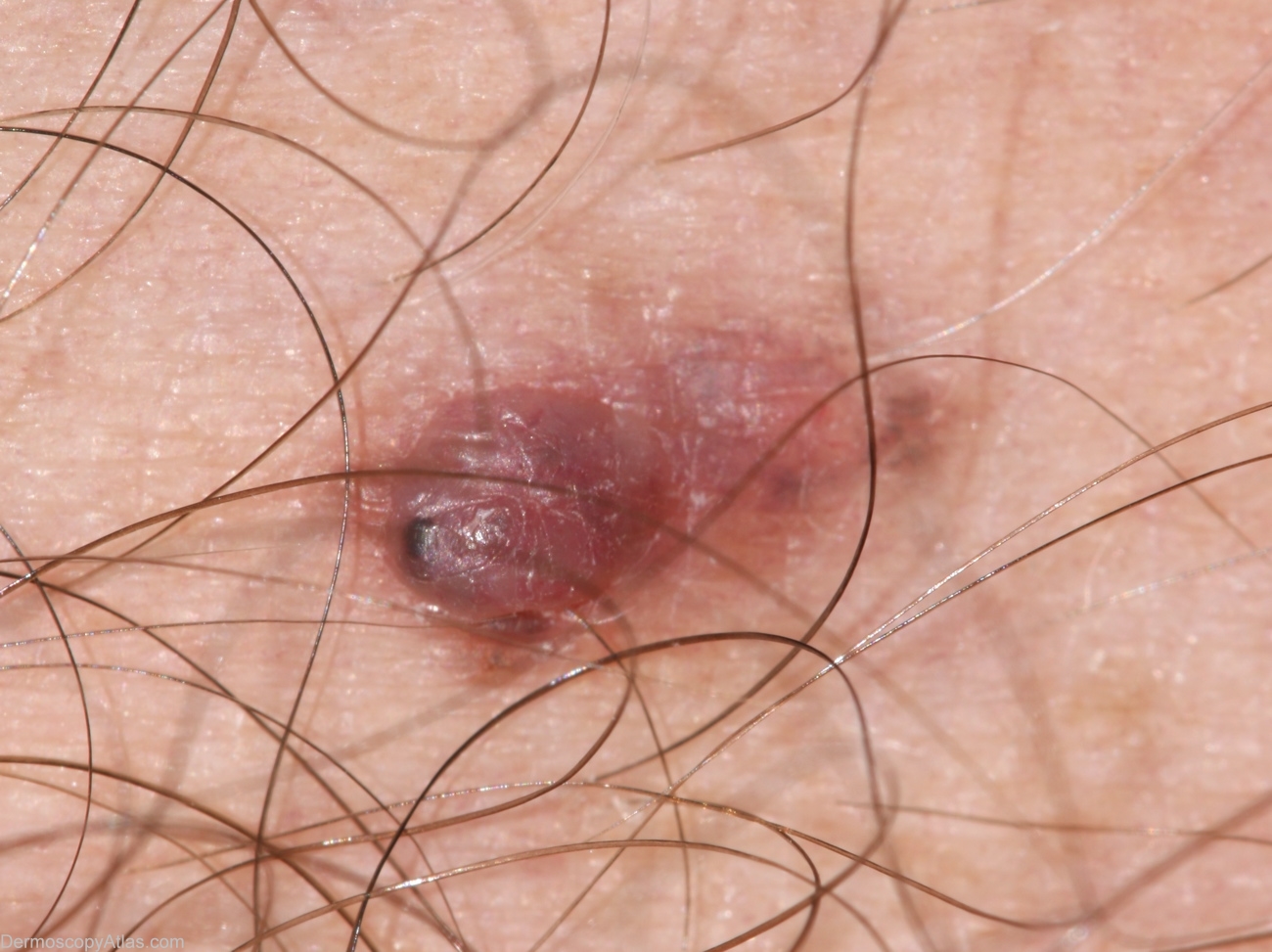
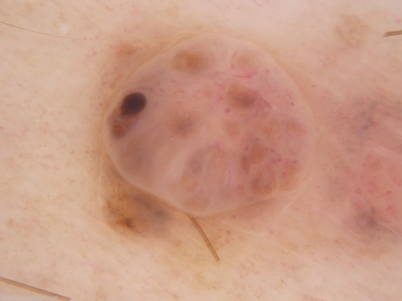
Description: The nodular component is not unlike a benign dermal naevus with maybe (in retrospect) more variability in vessel morphology and the unusual solitary brown clod. The macular component shows dots grey and more unusual fine hairpin vessels. Thes features suggest a (symptomatic) regressing melanocytic lesion.
History: 35 year old male with atypical mole syndrome. This lesion noted at examination prompted by melanoma 393. On asking, the patient mentioned this lesion had been irritated by clothing when active for the last 6-9 months. Histology reported as: Sections show a level 3 (0.8mm thick) nodular melanoma with prominent old regression. There is no ulceration, dermal mitoses or lymphocytic infiltrate.