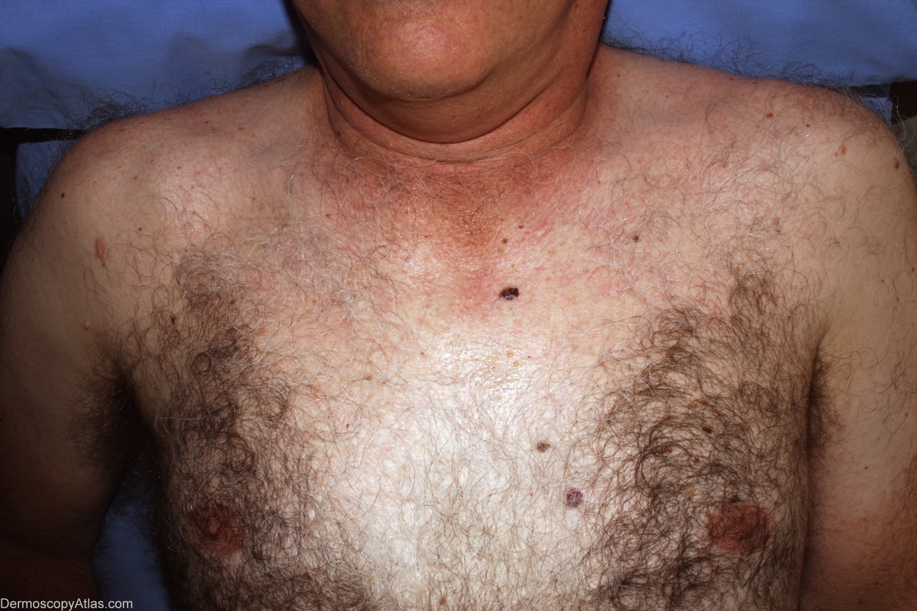
Loading
TeleDermConsult-logo
  • Recent Submissions
  • Search
  • Diagnosis
  • Quiz
  • Image comparison
  • Submit
  • Teaching Cases
  • Index
  • About

<< Back to Diagnosis Page

Image Number #2942 (Melanoma invasive)

Site: Chest

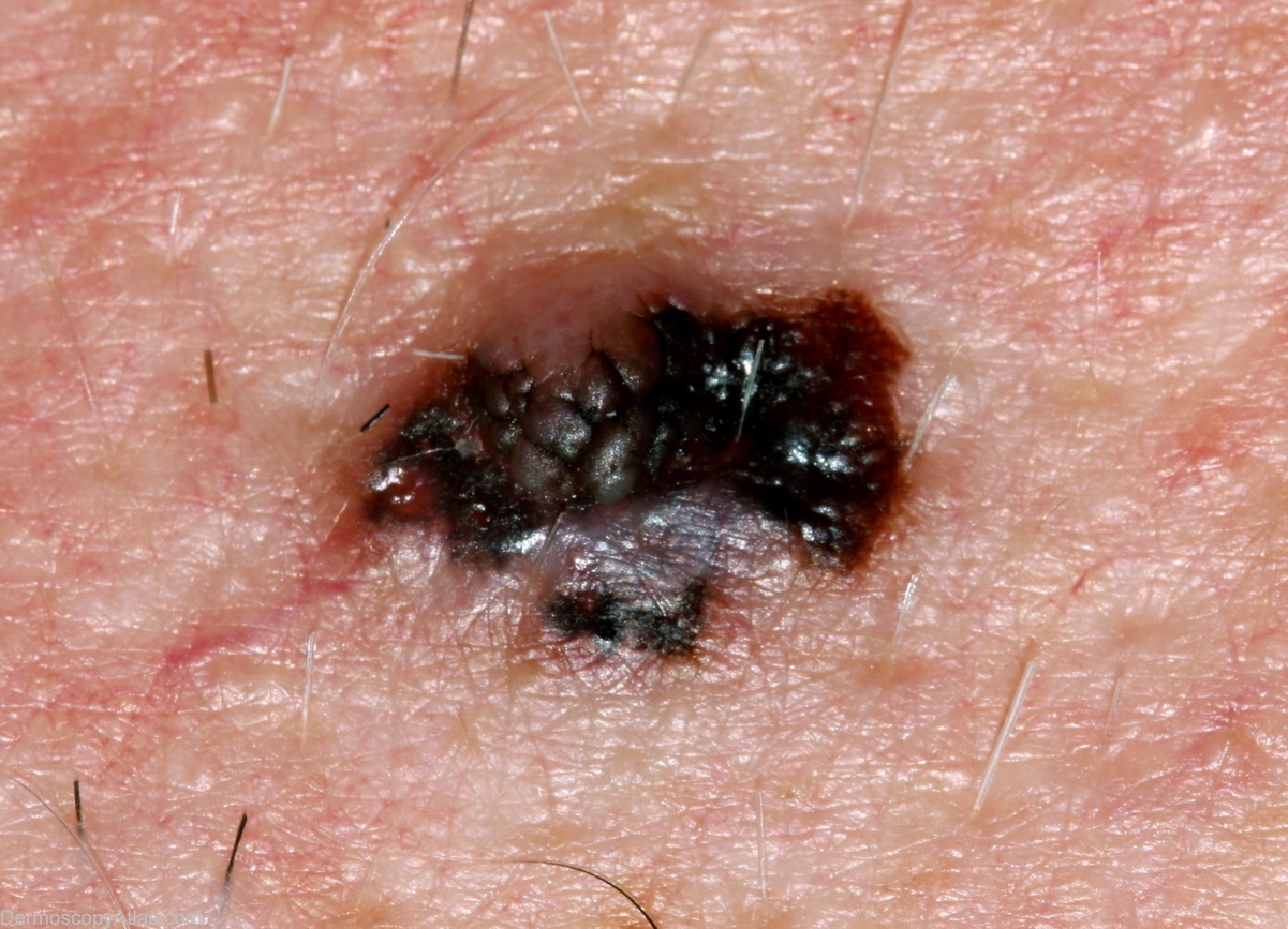
Diagnosis: Melanoma invasive

Sex: M

Age: 58

Type: Dermlite Non Polarised

Submitted By: Alan Cameron

View Full Size Image

Click Here to View Details

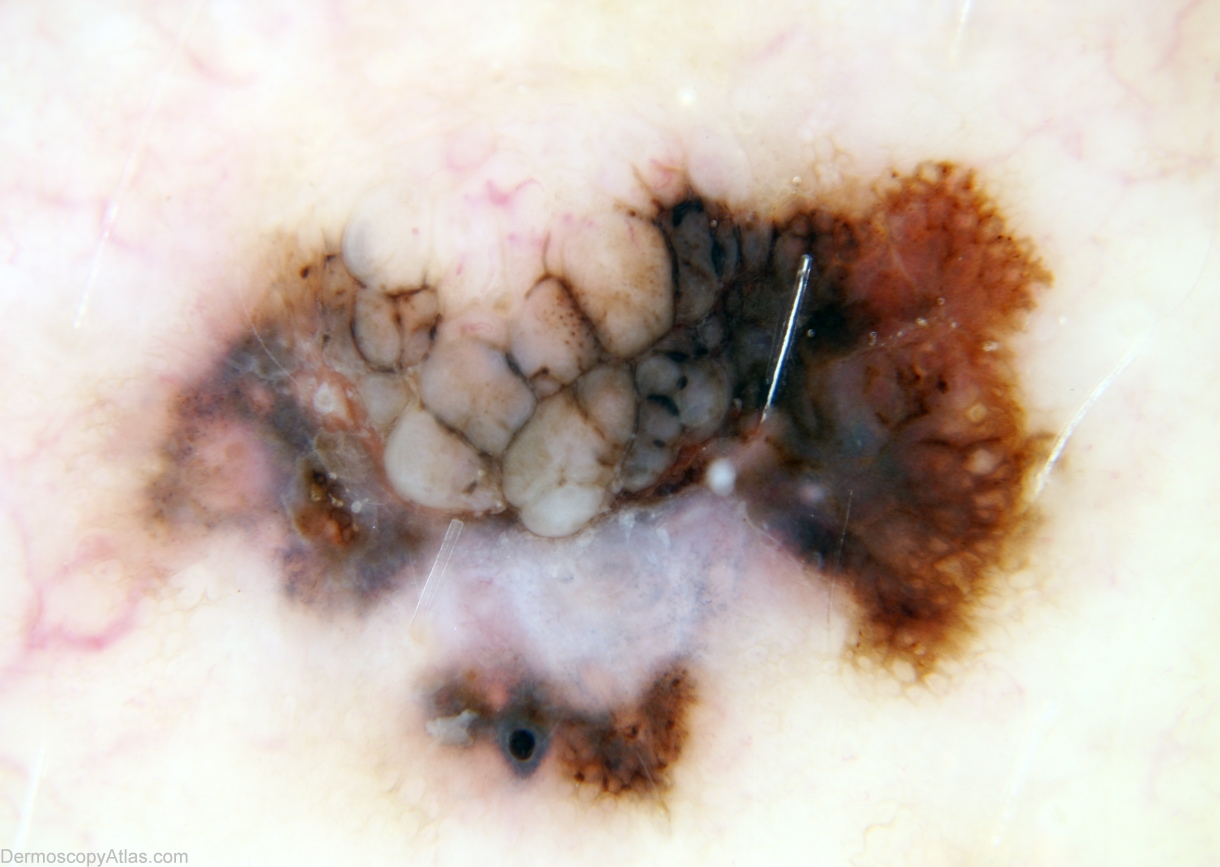
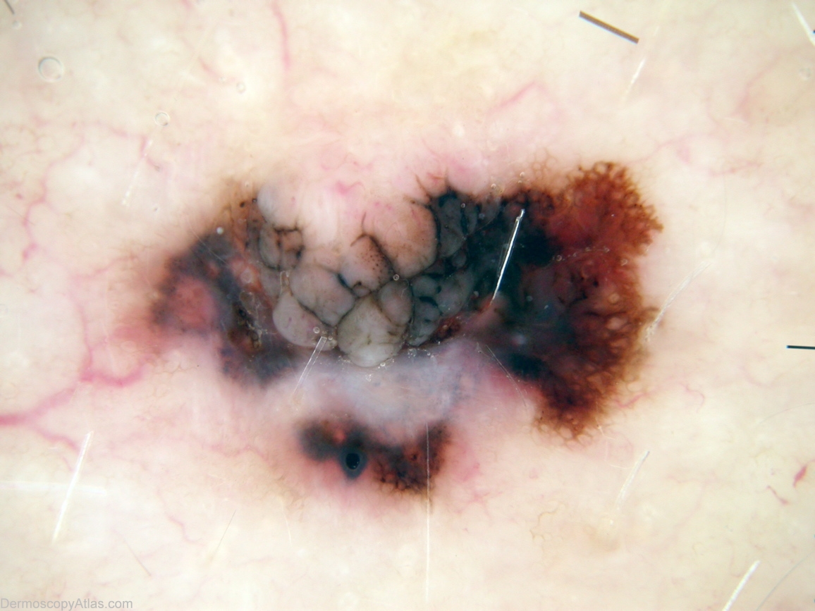
Description: Macroscopically this can only be melanoma.

History: 58 year old man, no past history but multiple atypical naevi. Patient considered this lesion to be longstanding and not changing, referred by his GP for assessment. Histology reported as; Sections show a Level 2 (0.5mm thick) superficial spreading melanoma arising in a benign intradermal naevus. There is no ulceration or dermal mitoses. There is a heavy lymphocytic infiltrate with prominent old regression. Reported by Dr David Weedon

DermNetNZ   eMedicine   PubMed   Dermatology Online   Archives   JAAD for "Melanoma invasive"

  • Recent Submissions
  • Search
  • Diagnosis
  • Quiz
  • Image comparison
  • Submit an Image
  • Teaching Cases
  • Index
  • About

Copyright 2022. Dermoscopy Atlas. ALL Rights Reserved.

Change Image