
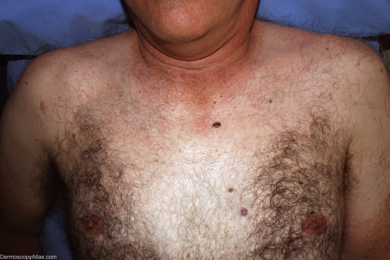
Site: Chest
Diagnosis: Melanoma invasive
Sex: M
Age: 58
Type: Dermlite Non Polarised
Submitted By: Alan Cameron
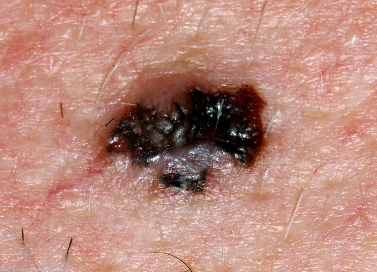
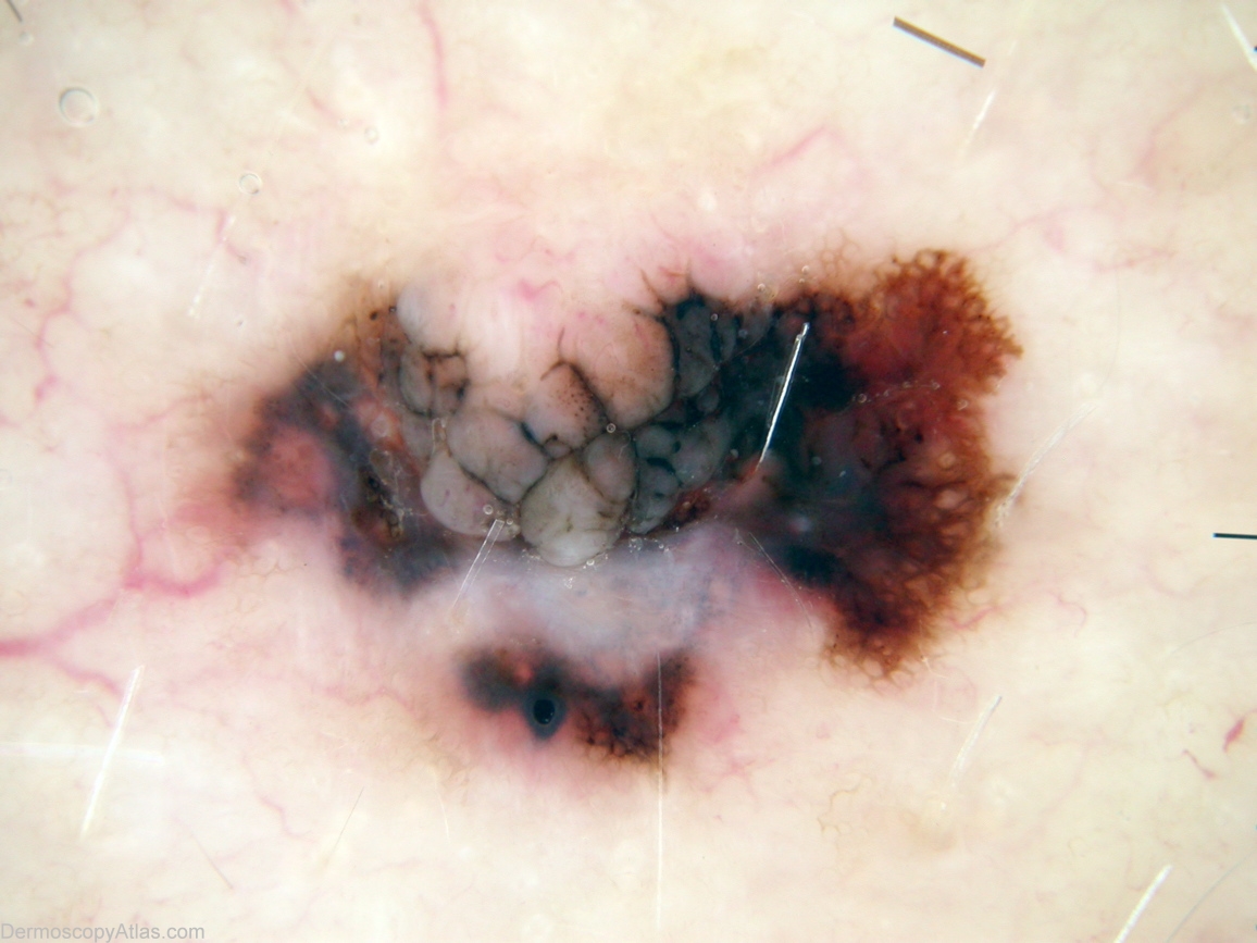
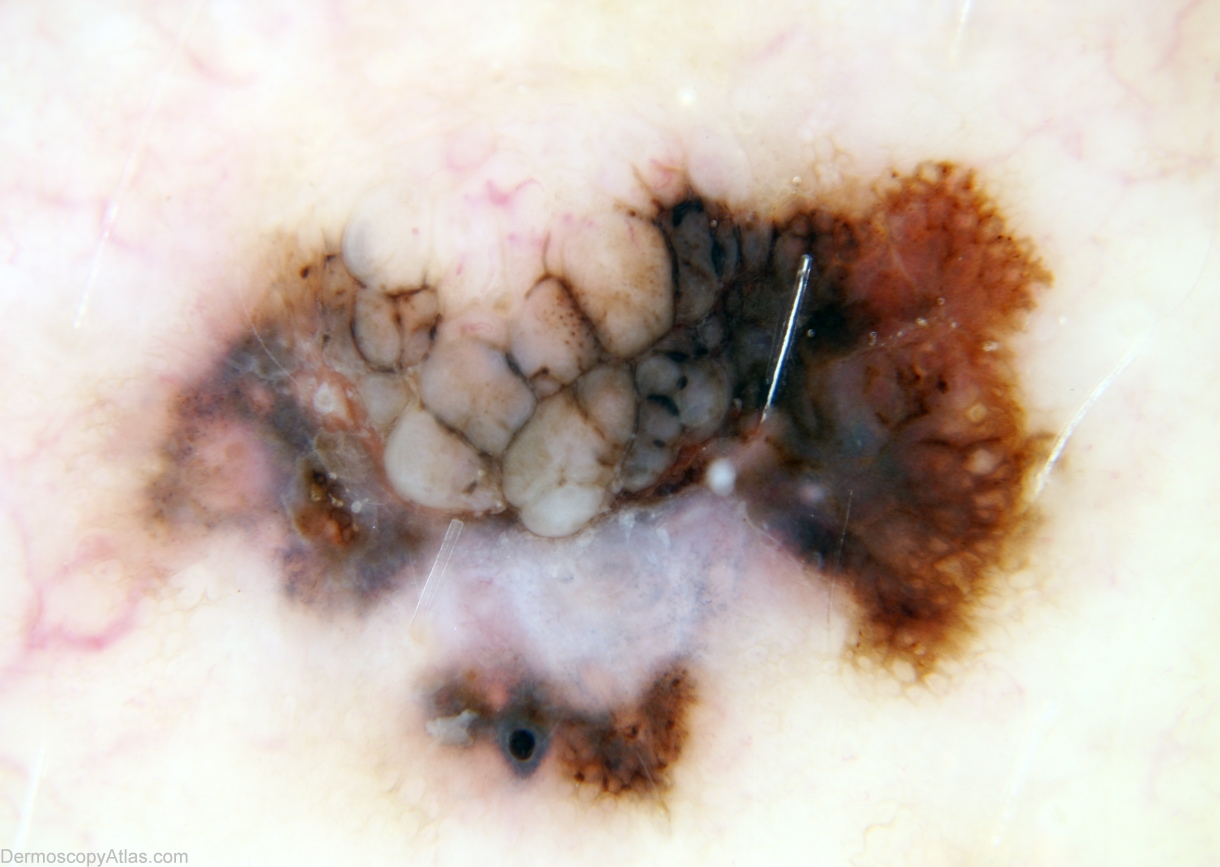
Description: Pattern is lines reticular thick and thin with segmental lines radial(top right), brown dots and structureless white. The intradermal naevus can be clearly seen top middle with brown and black lines demarcating sections of naevus, and brown dots in one section. Professor Weedon comments that melanoma barely involves this intradermal naevus, having skirted all around it. White 'clod' inferior to bright hair is artefact.
History: 58 year old man, no past history but multiple atypical naevi. Patient considered this lesion to be longstanding and not changing, referred by his GP for assessment. Histology reported as; Sections show a Level 2 (0.5mm thick) superficial spreading melanoma arising in a benign intradermal naevus. There is no ulceration or dermal mitoses. There is a heavy lymphocytic infiltrate with prominent old regression. Reported by Dr David Weedon