
Site: Abdomen
Diagnosis: Melanoma invasive
Sex: M
Age: 65
Type: Dermlite Non Polarised
Submitted By: Cliff Rosendahl
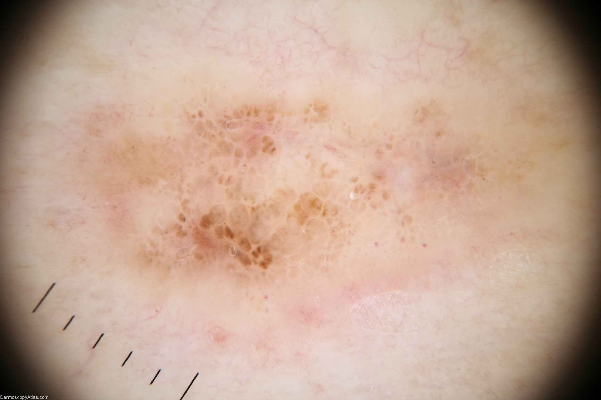
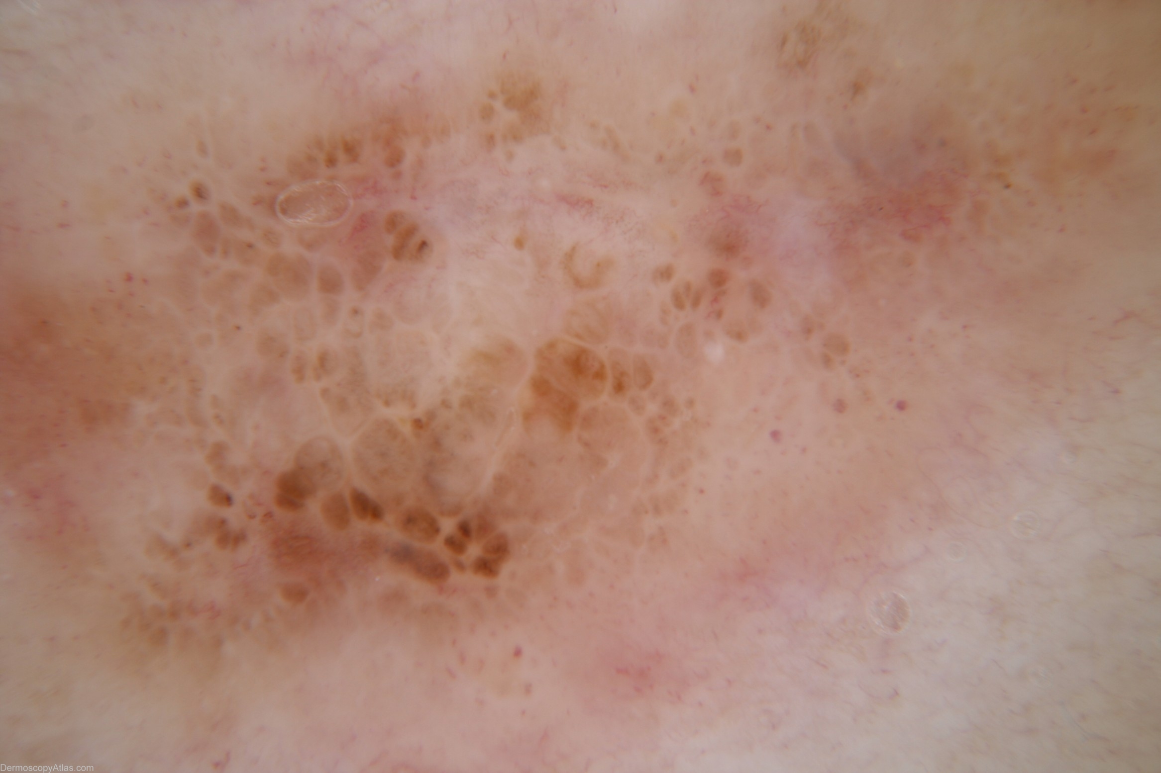
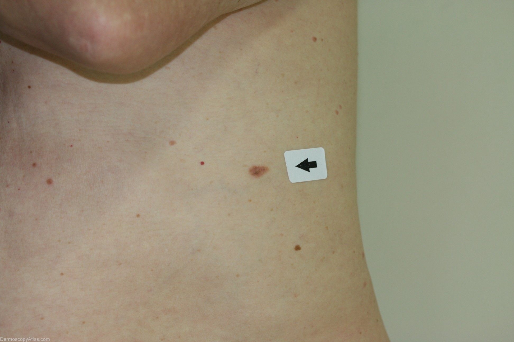
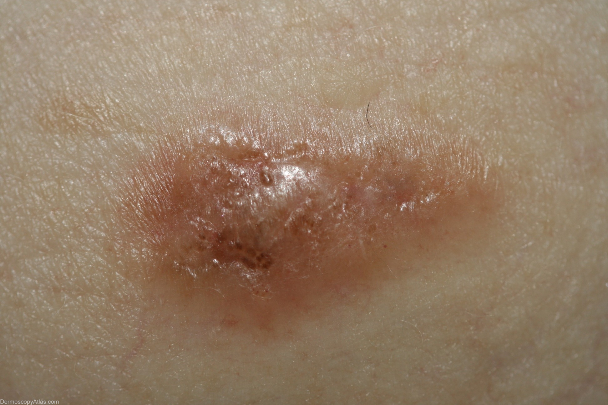
Description: Macro image - This shows an unusual lesion with a translucent quality asymmetry and more than one colour. It is also raised rather than macular and this meant that if there was any suspicion of melanoma monitoring would be contraindicated.
History: This gentleman with no previous history of melanomas presented for a skin examination and this lesion was noticed. I was unsure whether it was melanocytic so performed a punch biopsy. The report stated that it was a benign dysplastic naevus. Because it was melanocytic an excision biopsy was performed which was reported as a level 2 melanoma with a Breslow thickness of 0.7mm. A third procedure was then necessary to achieve appropriate margins.