
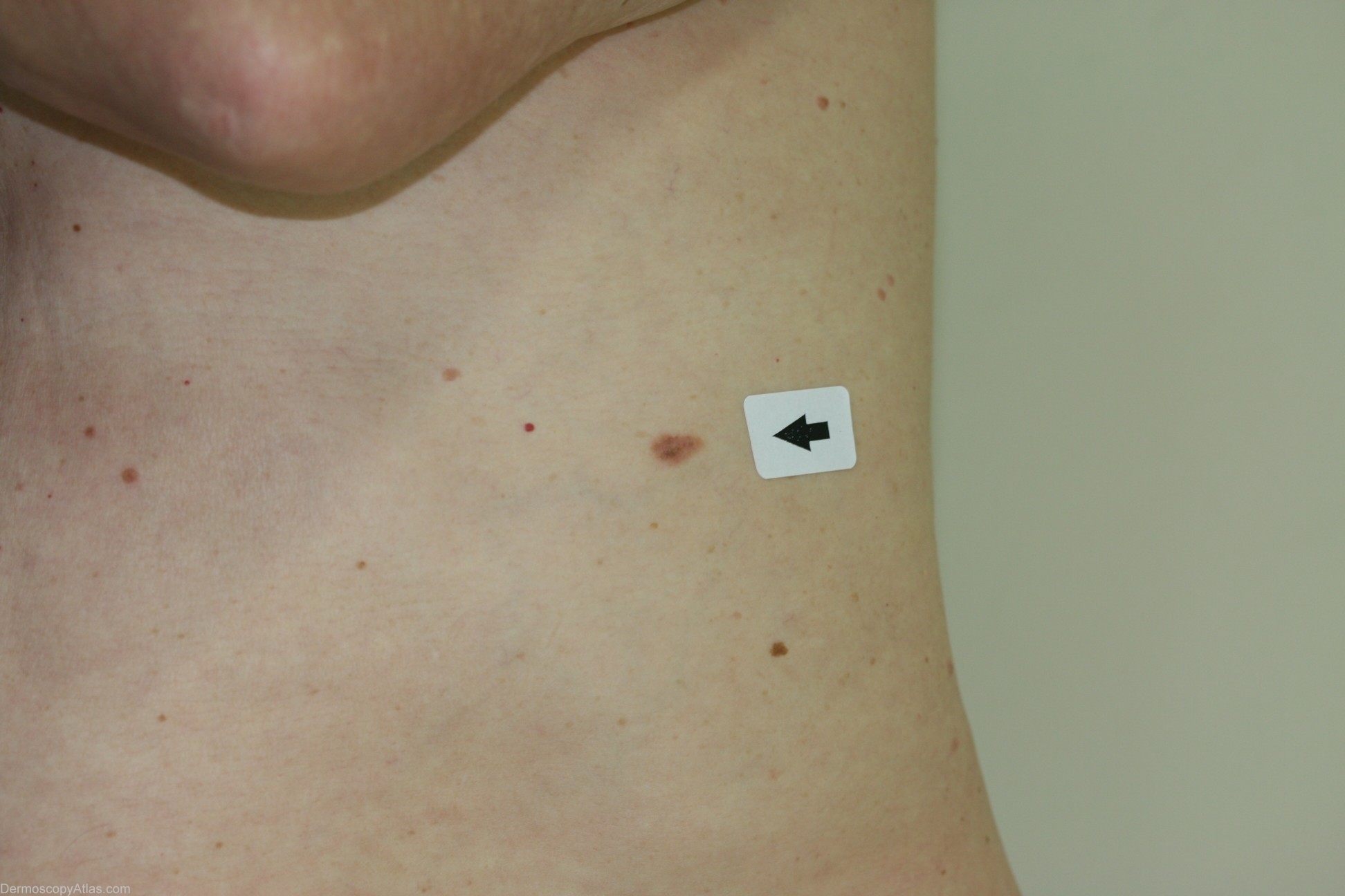
Site: Abdomen
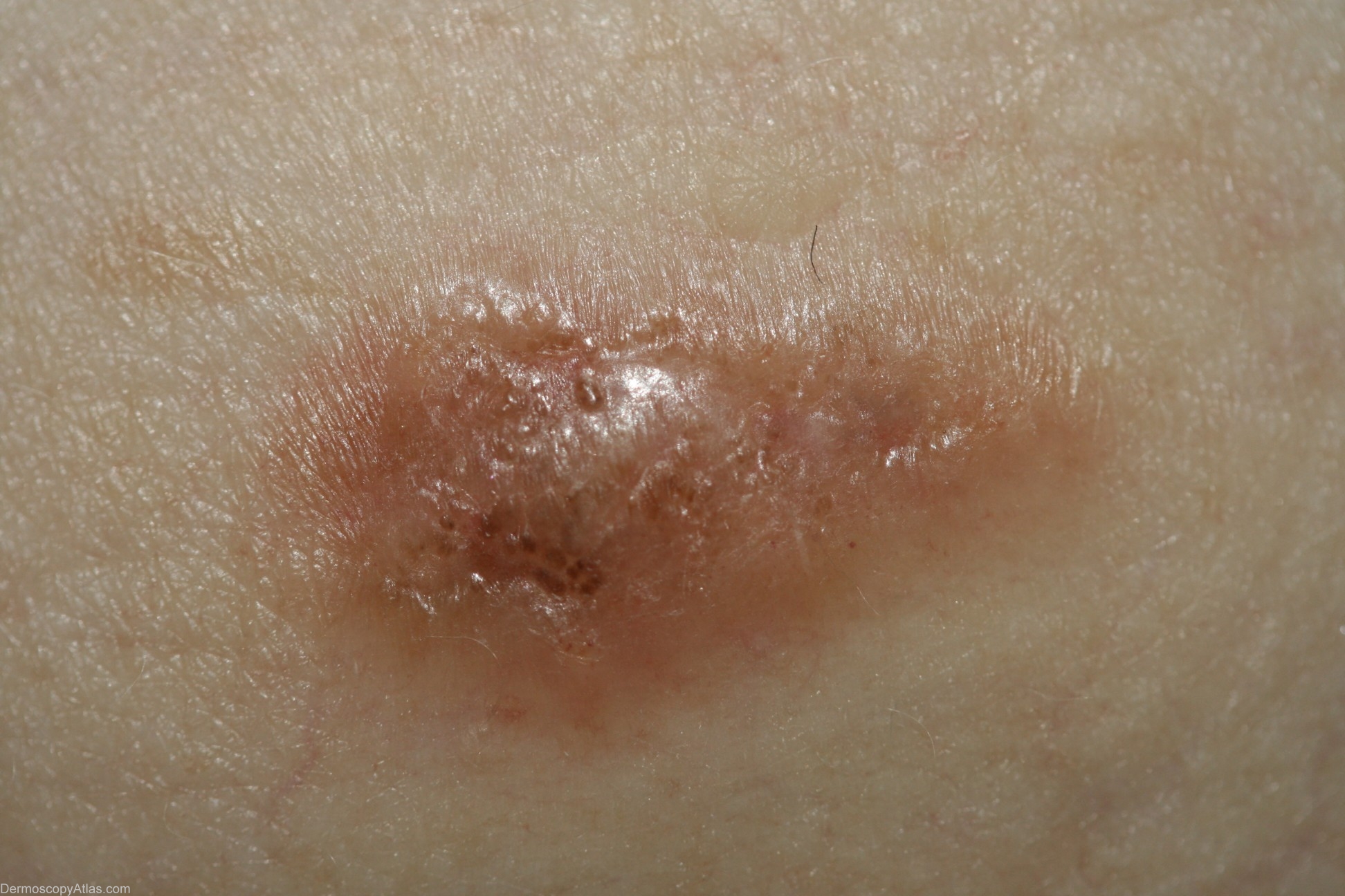
Diagnosis: Melanoma invasive
Sex: M
Age: 65
Type: Dermlite Non Polarised
Submitted By: Cliff Rosendahl
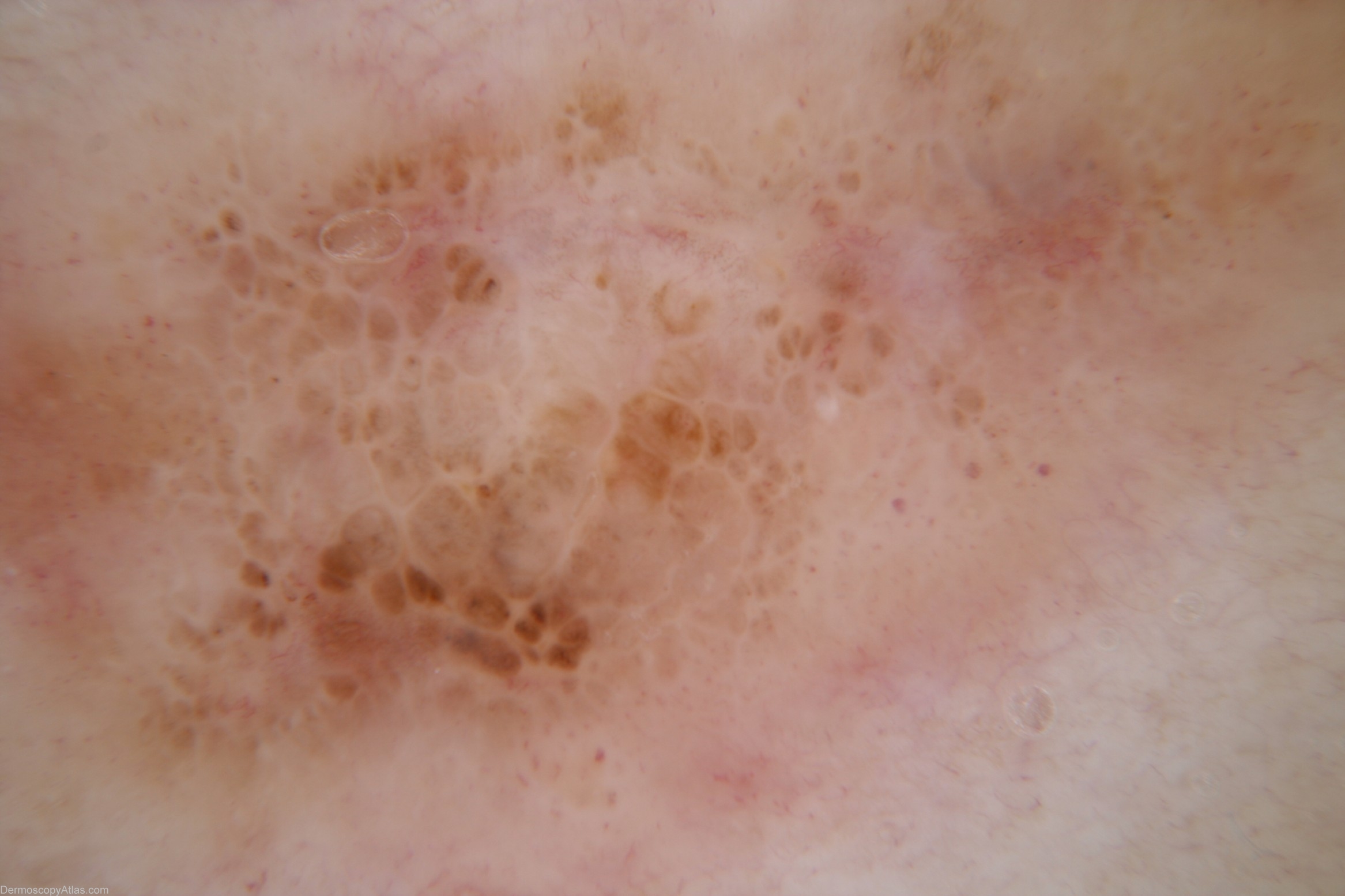
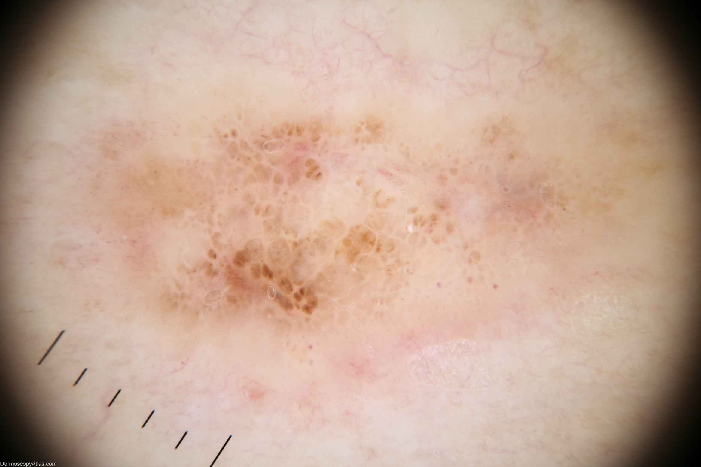
Description: Dermoscopy image Pattern - Clods, arguably white lines reticular, structureless area, structureless area. There is an element of chaotic asymmetry in the arrangement of clods Colours - Light brown, dark brown, red, grey Clues to melanoma - focal grey colour, white lines reticular, dot vessels
History: This gentleman with no previous history of melanomas presented for a skin examination and this lesion was noticed. I was unsure whether it was melanocytic so performed a punch biopsy. The report stated that it was a benign dysplastic naevus. Because it was melanocytic an excision biopsy was performed which was reported as a level 2 melanoma with a Breslow thickness of 0.7mm. A third procedure was then necessary to achieve appropriate margins.