
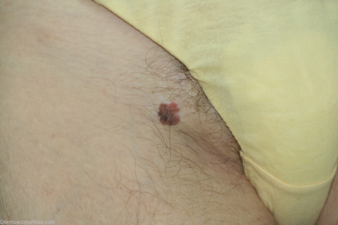
Site: Thigh
Diagnosis: Pigmented Intraepidermal carcinoma
Sex: F
Age: 69
Type: Dermlite Non Polarised
Submitted By: Cliff Rosendahl
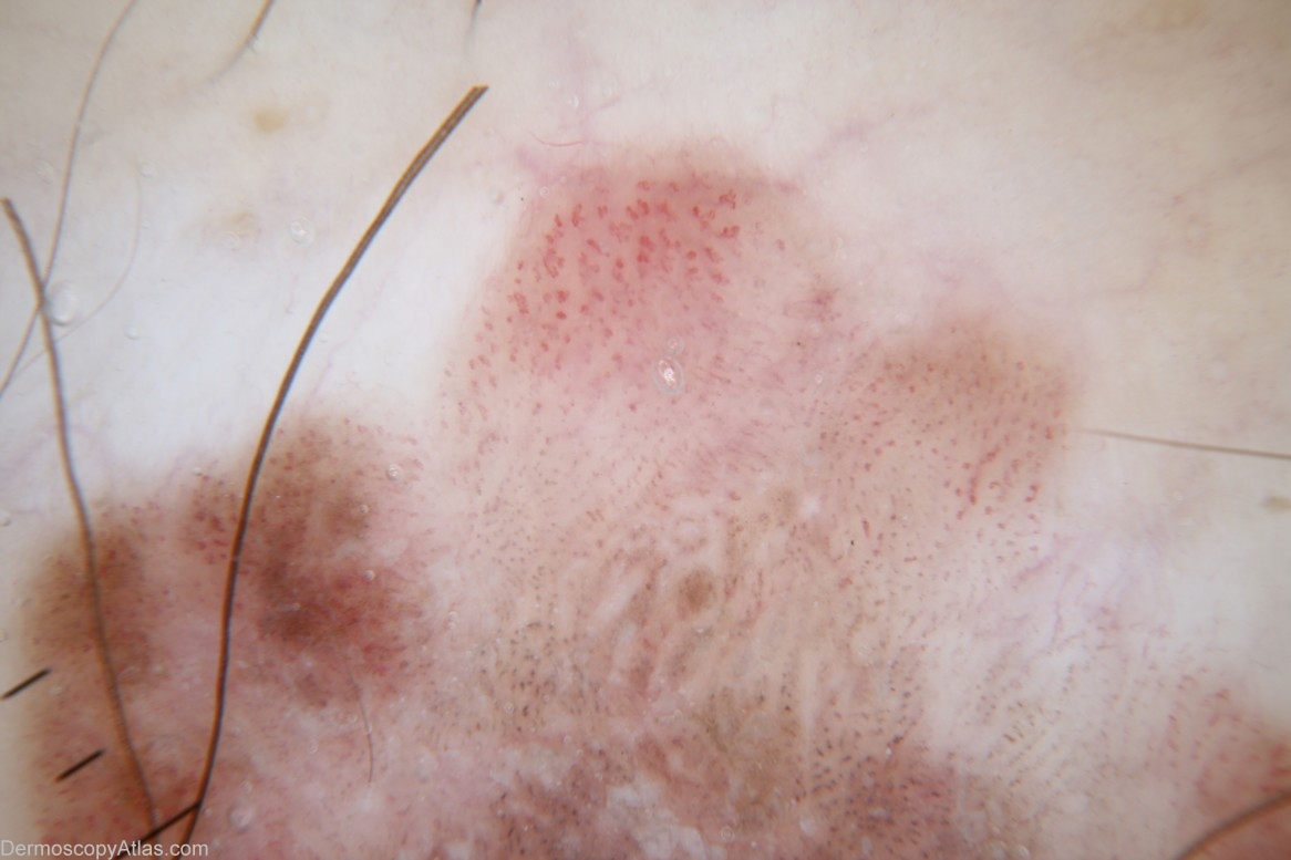
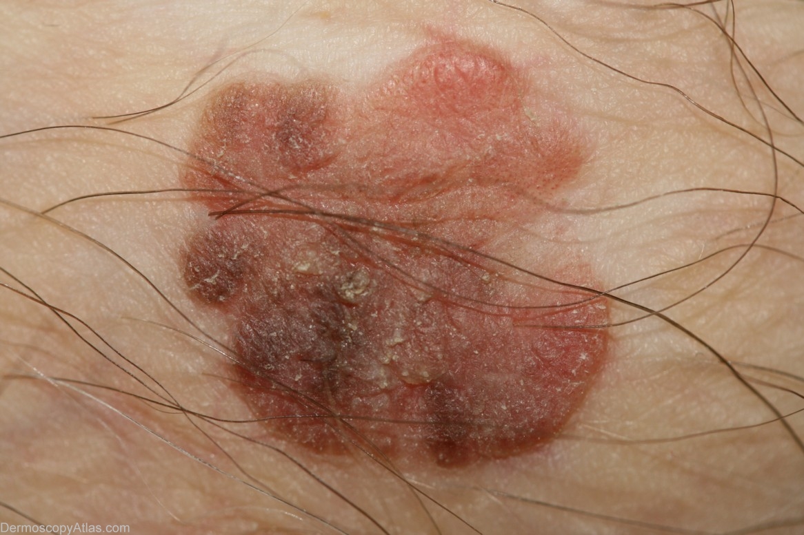
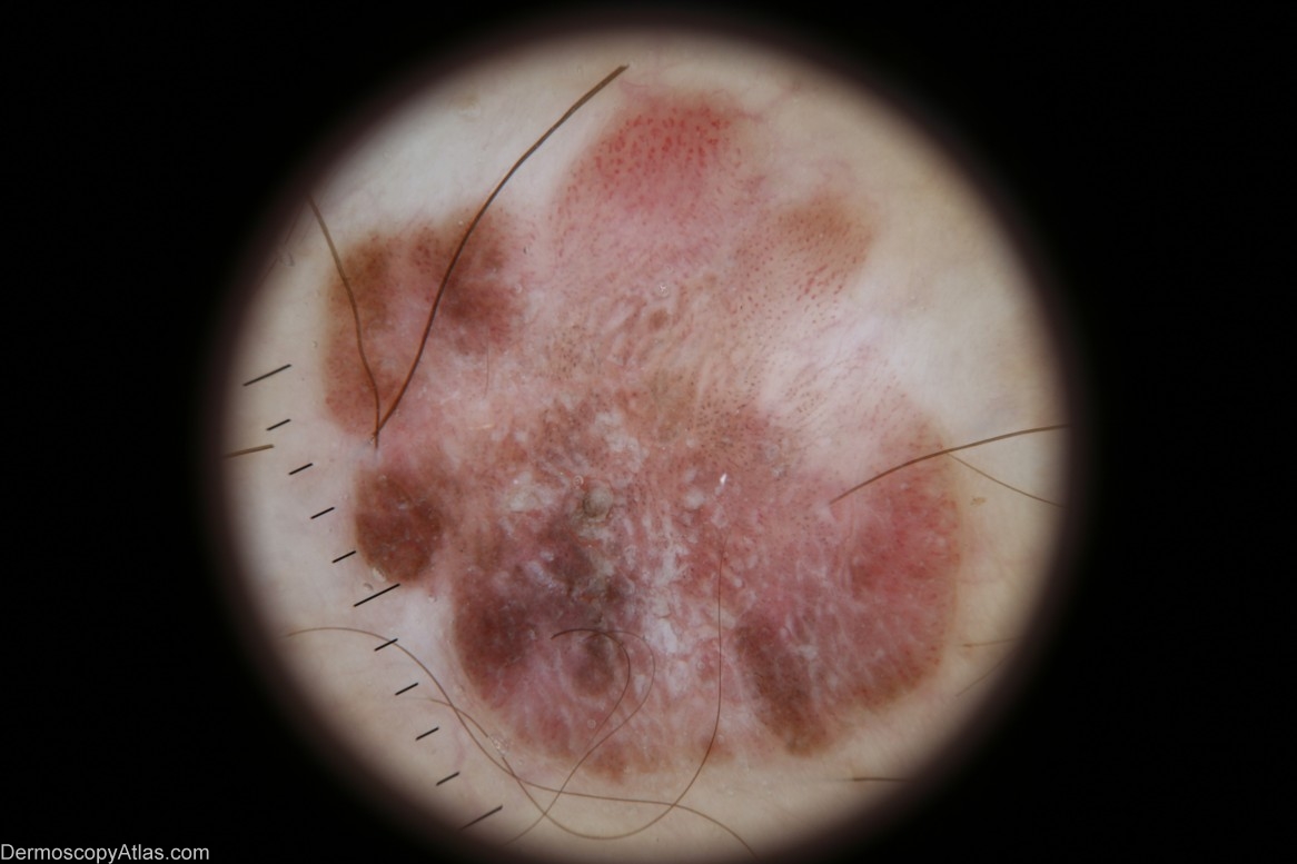
Description: There is no pigment network but this lesion can be described as "melanocytic by default".The lesion is asymmetric and has several colours including brown,red and white. There is a cluster of glomerular vessels neatly arranged. There is a cluster of dot vessels also neatly aligned in rows. There are areas of amorphous brown pigmenent. There are brown dots. There is a large amount of scar-like depigmentation. This was a pigmented IEC but melanoma was on the differential diagnosis.
History: This lesion was encountered at a routine skin check. The patient stated that it had been present for the last 4 years.