
Site: Knee
Diagnosis: Melanoma nodular
Sex: F
Age: 67
Type: Heine
Submitted By: Ian McColl
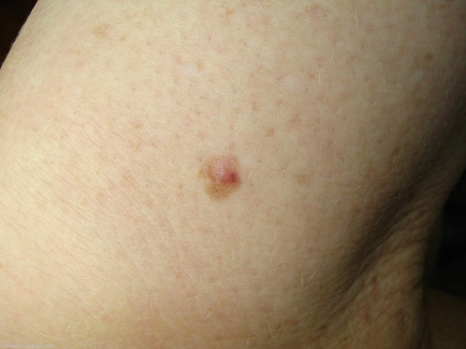
Description: The clinical image of the thigh lesion. The peripheral pigmented lentigene like lesion had been present for some time but the pink nodule was new. It does show a small fleck of pigmentation.
History: This lesion had changed recently. What do you think it is and do you feel that the information available dermatoscopically was enhanced by using the Automatic light level toggle (single click only) in my Image management programme?
The path report of the initial excision was " A superficial spreading malignant melanoma . The bulk of the melanoma is Level 1 (in situ). There is however a central dermally invasive component ( Level 3, 0.80mm in greatest depth). No tumour infiltrating lymphocytes, no regression."
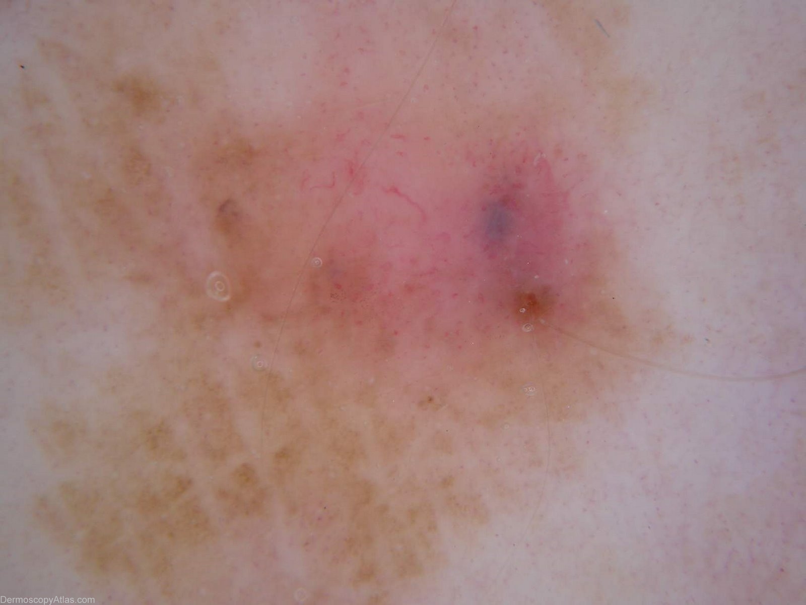
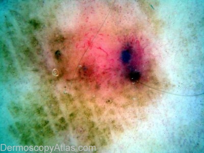
You have a pink papule arising in a background pigmented lesion. Essentially melanoma until proven otherwise. The dermatoscopy is interesting and to my view superficially misleading. On the non enhanced views it looks like a pigmented BCC but I felt the automatic colour adjust made the dermatoscopic diagnosis easier by highlighting the melanin and the polymorphous vessels.
View the Blog discussion of this case