
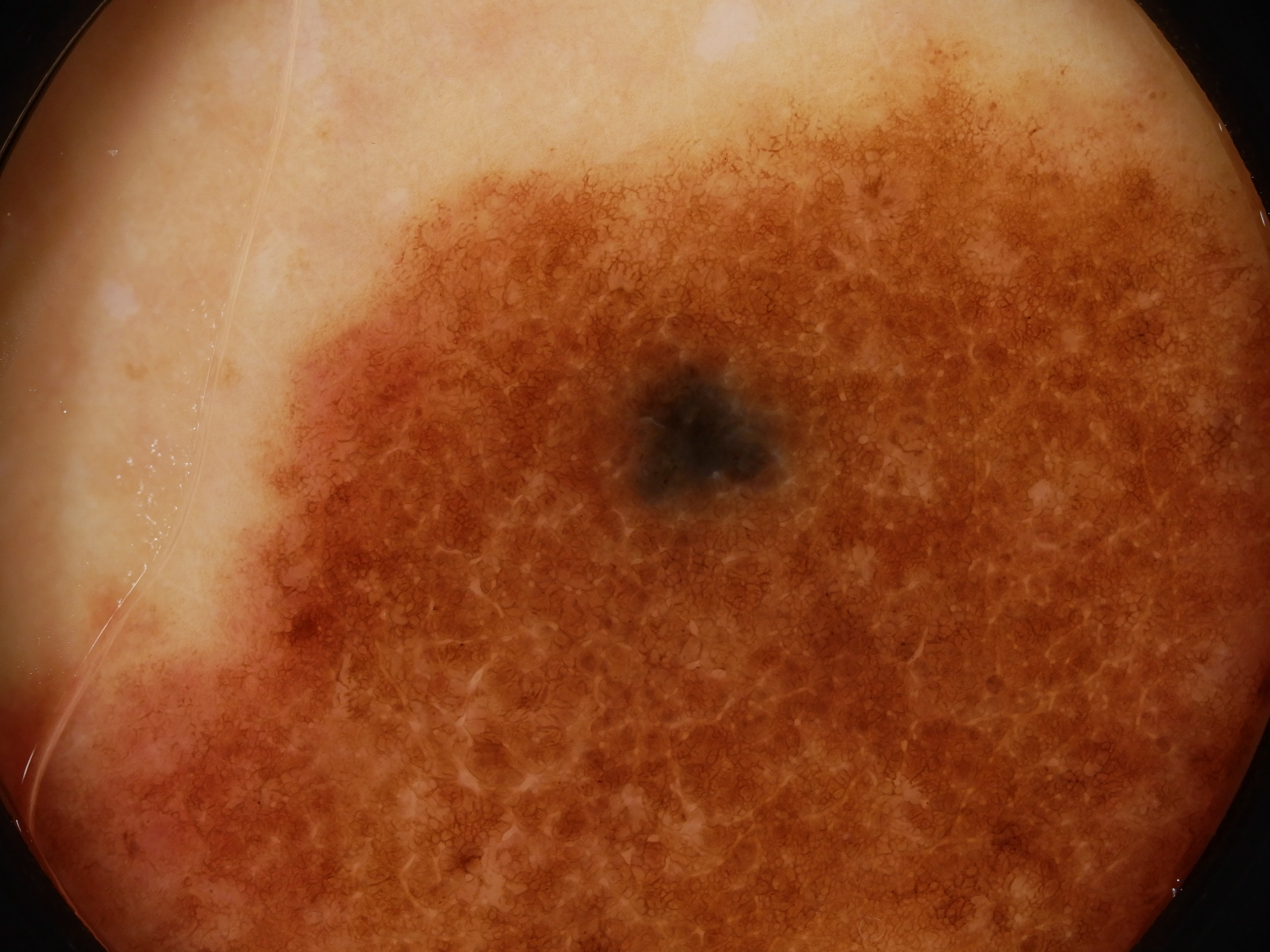
Site: Leg
Diagnosis: Congenital nevus
Sex: F
Age: 35
Type: Heine
Submitted By: Ian McColl
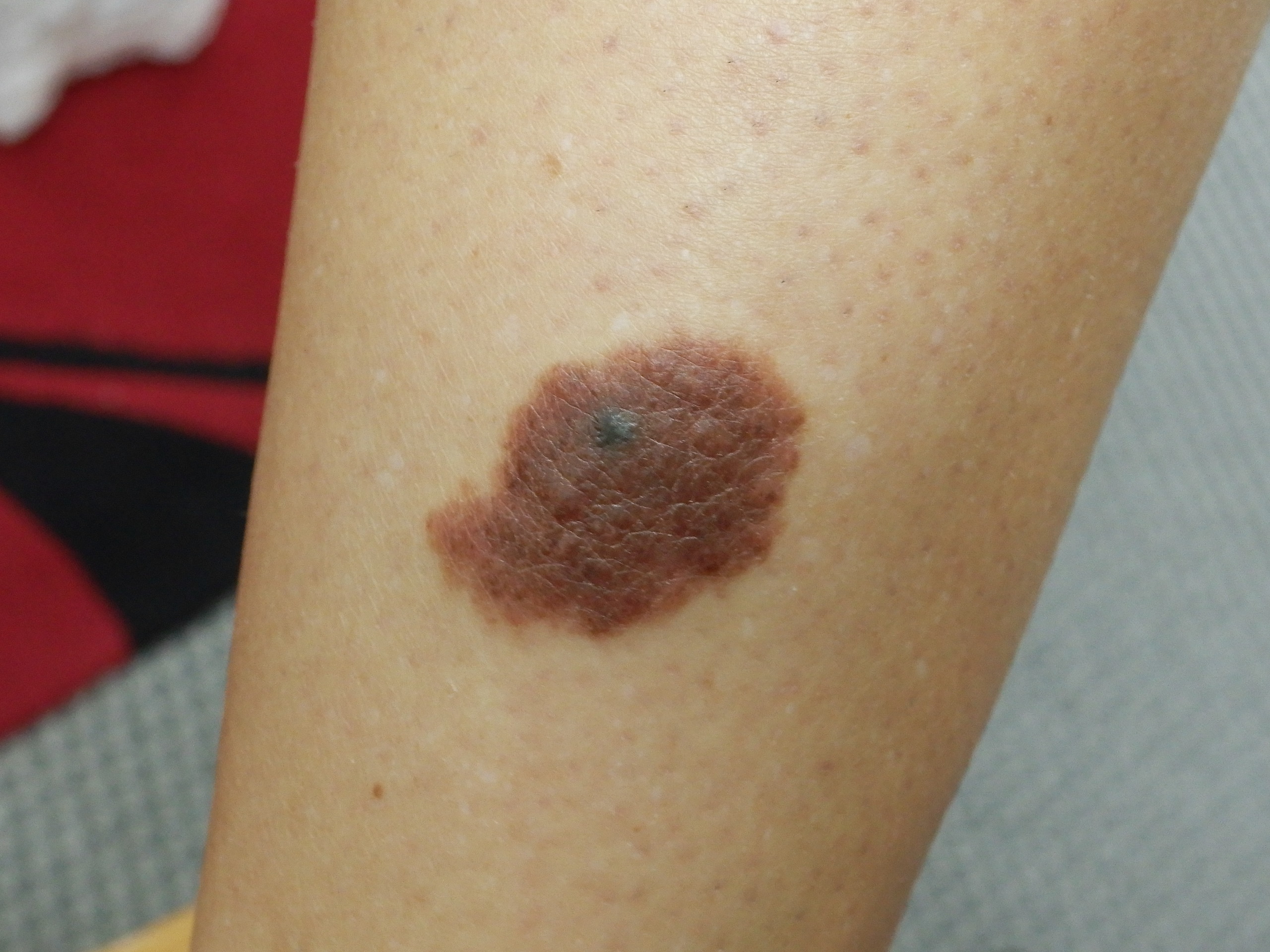
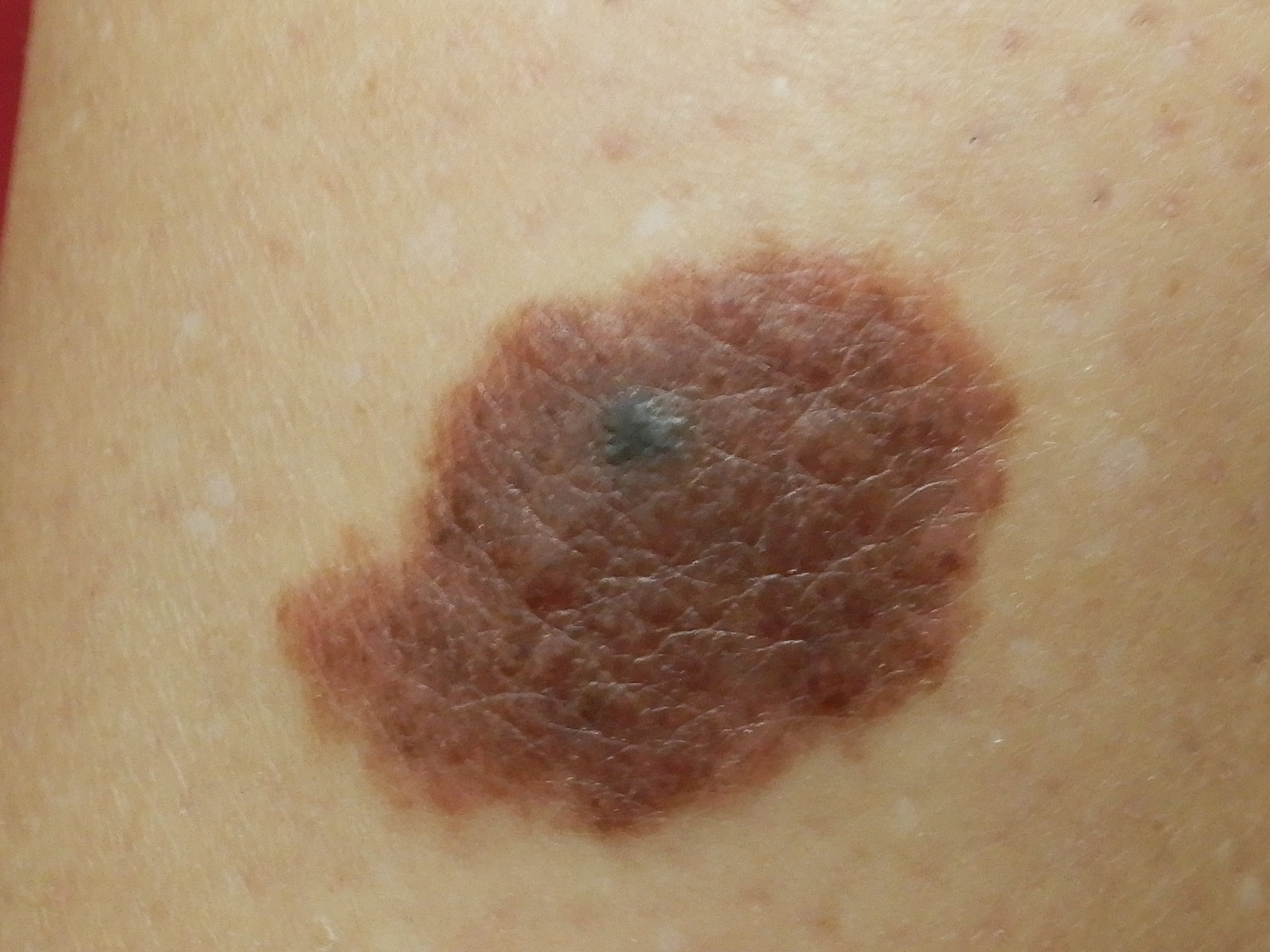
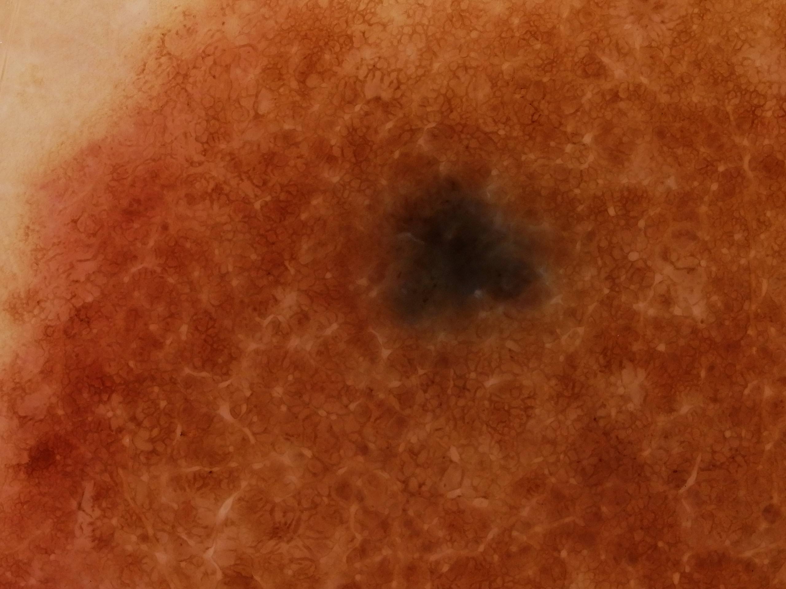
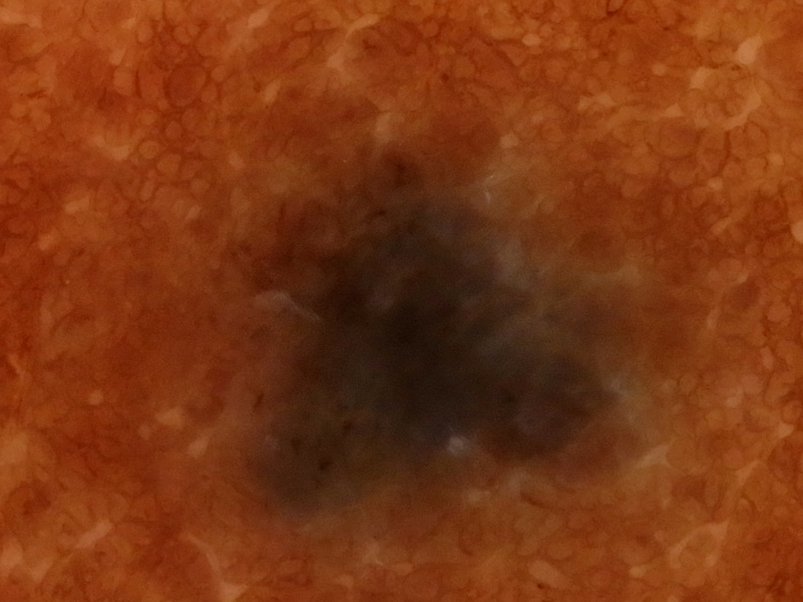
Description: Dermatoscopy
History:
A 35 year old female noted this darker area change in her congenital nevus over a period of 6 months. What would you do? Reassure, Excise the whole nevus or partial biopsy or completely excise just the darker pigmented area? What do you think it is? How would you approach this case?
1 Excise the whole congenital nevus 7% (3 votes)
2 Partially biopsy the pigmented area 15% (6 votes)
3 Completely excise only the pigmented area 20% (8 votes)
4 Excise the pigmented area and part of the surrounding congenital nevus 59% (24 votes)
Conclusion This was a deep penetrating nevus . I excised it and a few mm of the surrounding congenital nevus .