Loading
TeleDermConsult-logo
  • Recent Submissions
  • Search
  • Diagnosis
  • Quiz
  • Image comparison
  • Submit
  • Teaching Cases
  • Index
  • About

<< Back to Diagnosis Page

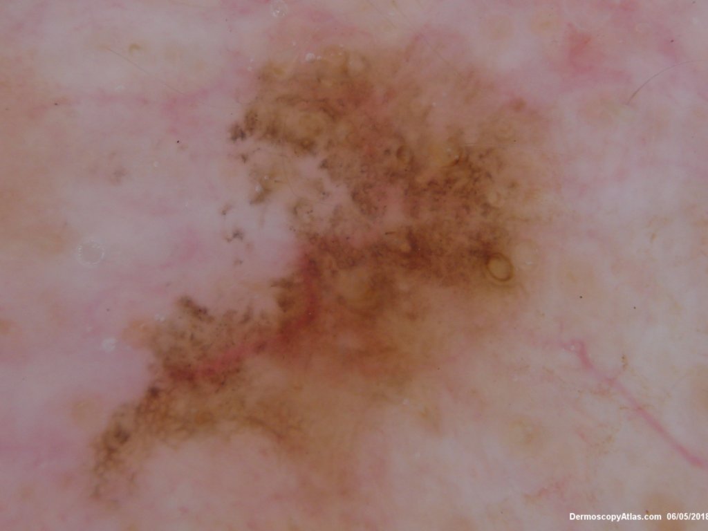
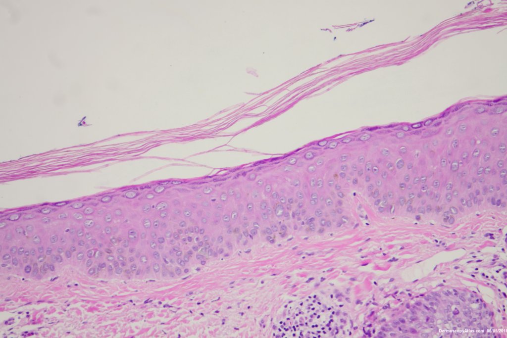
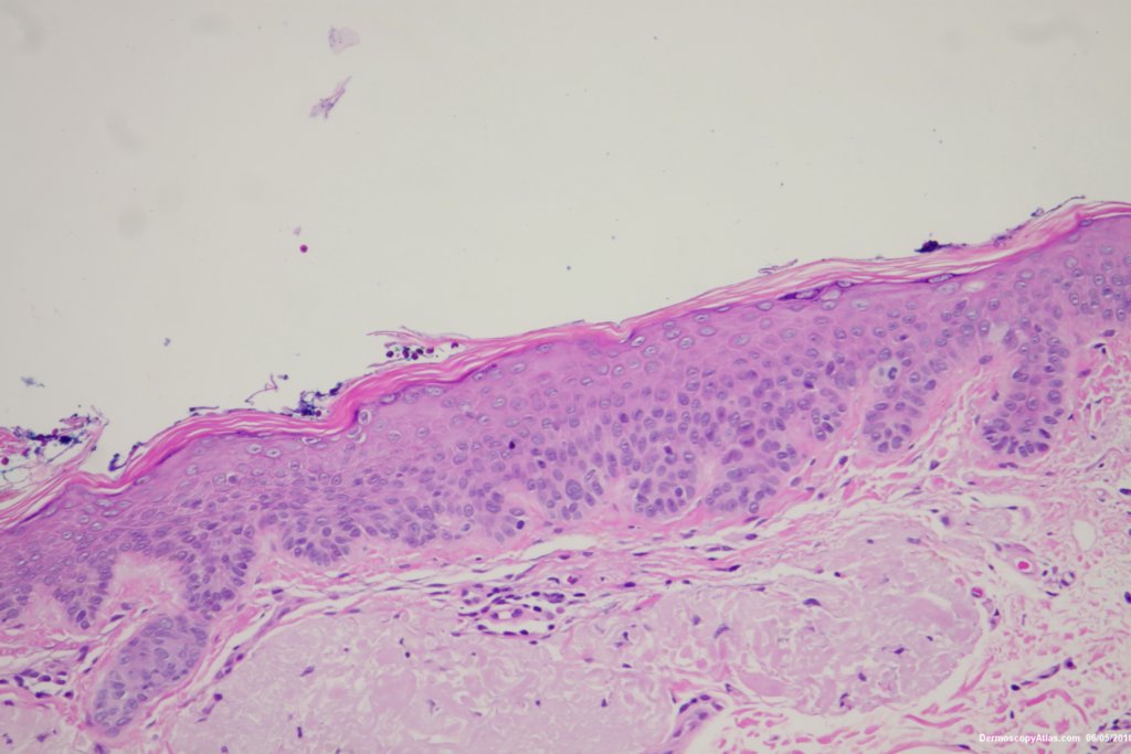
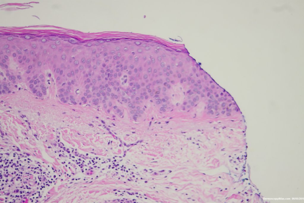
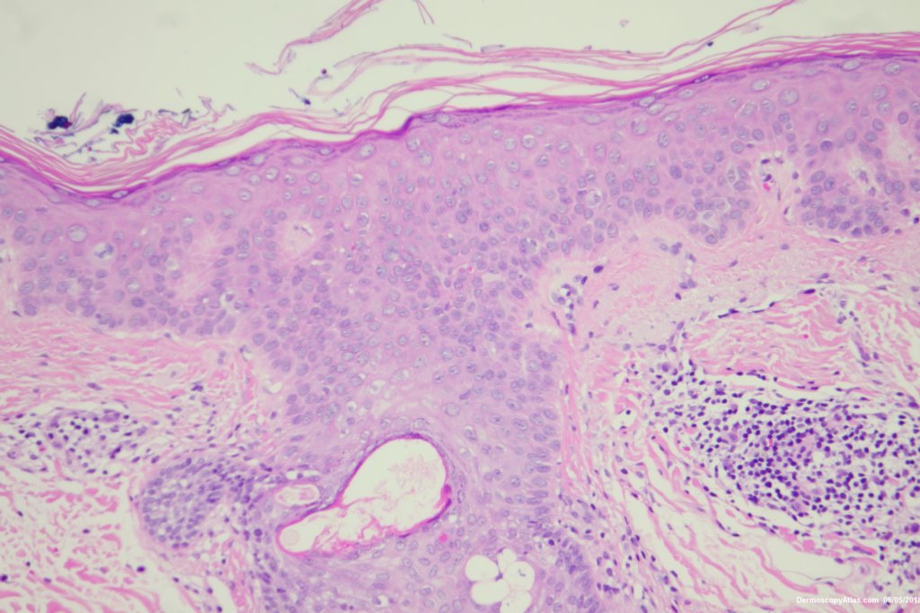
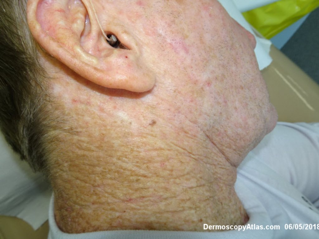
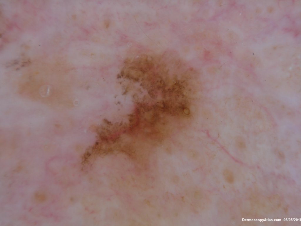
Image Number #3448 (Pigmented Intraepidermal carcinoma)

Site: Neck side

Diagnosis: Pigmented Intraepidermal carcinoma

Sex: M

Age: 67

Type: Dermlite Polarised

Submitted By: Ian McColl

View Full Size Image

Click Here to View Details

Description: Histology Pigmented IEC

History:

 Some pigmented lesions look melanocytic, However this is a pigmented intraepidermal carcinoma. Some areas show more full thickness atypia than others. The dermatoscopy shows some dots in rows but there are other grey dots showing regression at one edge.
DermNetNZ   eMedicine   PubMed   Dermatology Online   Archives   JAAD for "Pigmented Intraepidermal carcinoma"

  • Recent Submissions
  • Search
  • Diagnosis
  • Quiz
  • Image comparison
  • Submit an Image
  • Teaching Cases
  • Index
  • About

Copyright 2022. Dermoscopy Atlas. ALL Rights Reserved.

Change Image