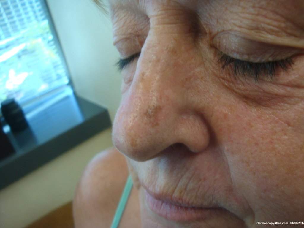
Loading
TeleDermConsult-logo
  • Recent Submissions
  • Search
  • Diagnosis
  • Quiz
  • Image comparison
  • Submit
  • Teaching Cases
  • Index
  • About

<< Back to Diagnosis Page

Image Number #3406 (Lentigo Maligna)

Site: Nose

Diagnosis: Lentigo Maligna

Sex: F

Age: 70

Type: Dermlite Polarised

Submitted By: Ian McColl

View Full Size Image

Click Here to View Details

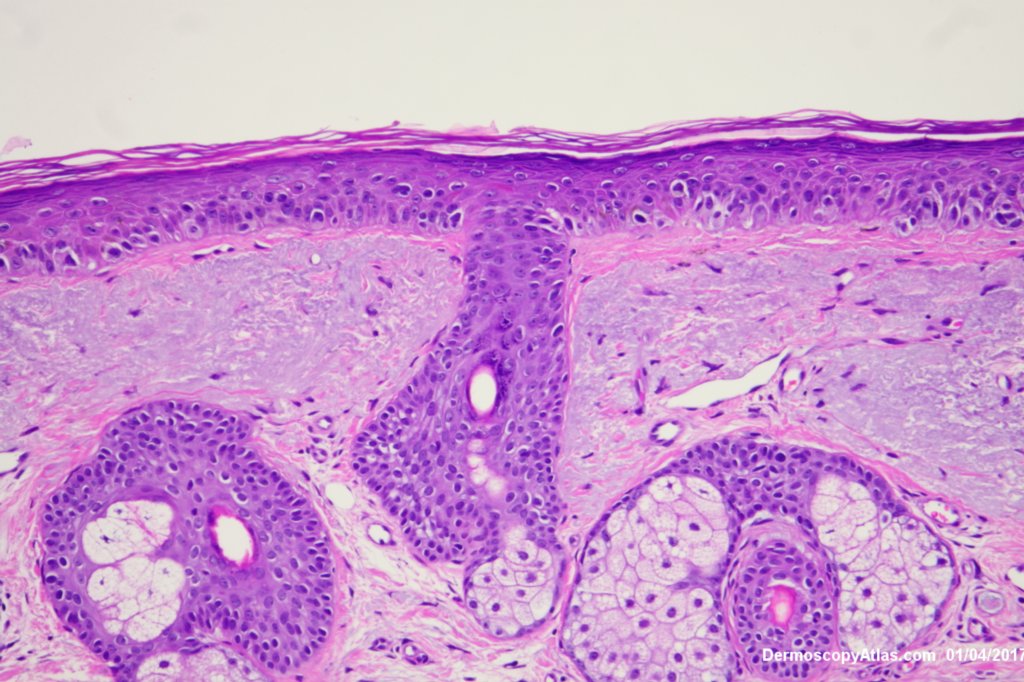
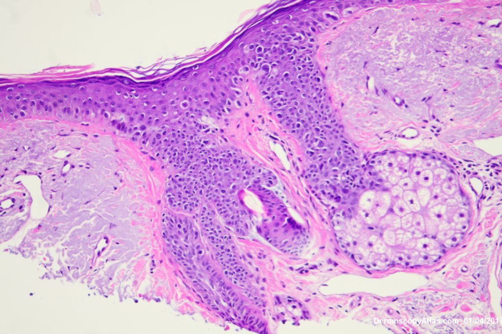
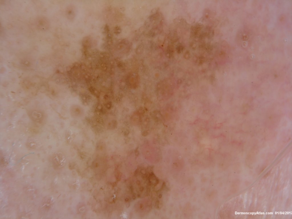
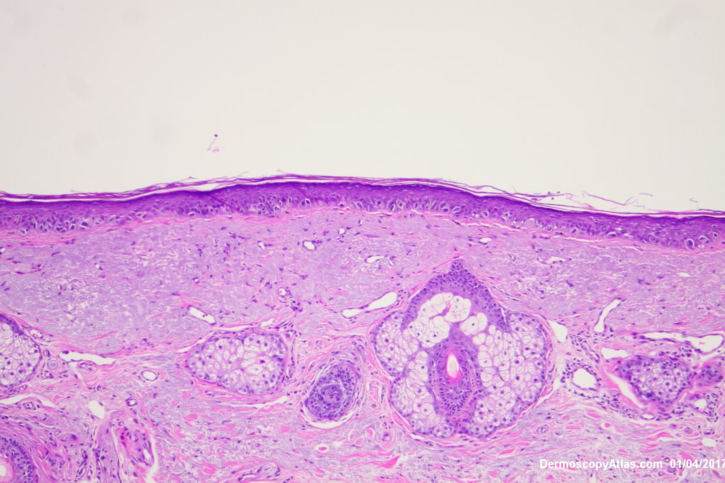
Description: Lentiginous proliferation of atypical melanocytes

History:

View a video of this case

This lesion had slowly grown over 6 months. No PH of non melanoma skin cancers or melanoma . 

The Dermatoscopy shows partial grey circles and the histology shows a lentiginous proliferation of atypical melanocytes involving the hair follicles.

Reported as lentigo maligna. Excised with a graft.

View this virtual slide of Lentigo Maligna

DermNetNZ   eMedicine   PubMed   Dermatology Online   Archives   JAAD for "Lentigo Maligna"

  • Recent Submissions
  • Search
  • Diagnosis
  • Quiz
  • Image comparison
  • Submit an Image
  • Teaching Cases
  • Index
  • About

Copyright 2022. Dermoscopy Atlas. ALL Rights Reserved.

Change Image