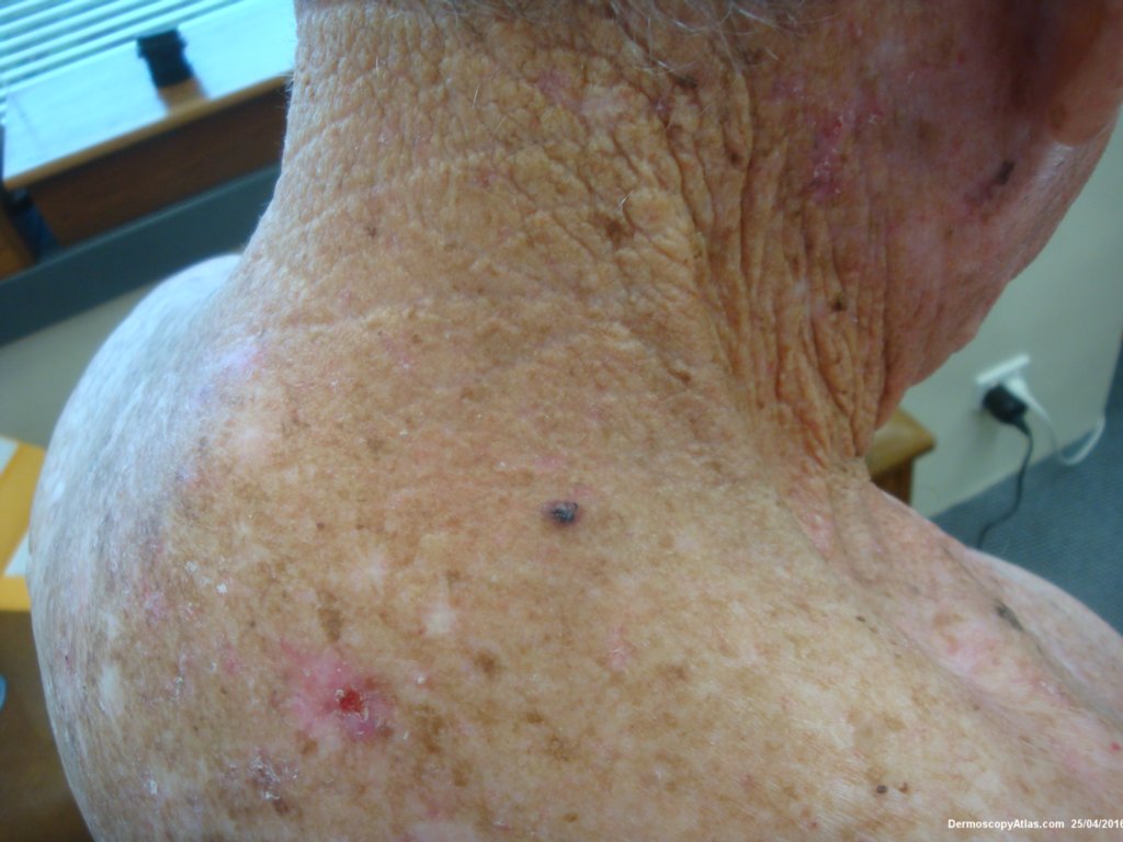
Loading
TeleDermConsult-logo
  • Recent Submissions
  • Search
  • Diagnosis
  • Quiz
  • Image comparison
  • Submit
  • Teaching Cases
  • Index
  • About

<< Back to Diagnosis Page

Image Number #3399 (BCC pigmented)

Site: Back

Diagnosis: BCC pigmented

Sex: M

Age: 78

Type: Dermlite Polarised

Submitted By: Ian McColl

View Full Size Image

Click Here to View Details

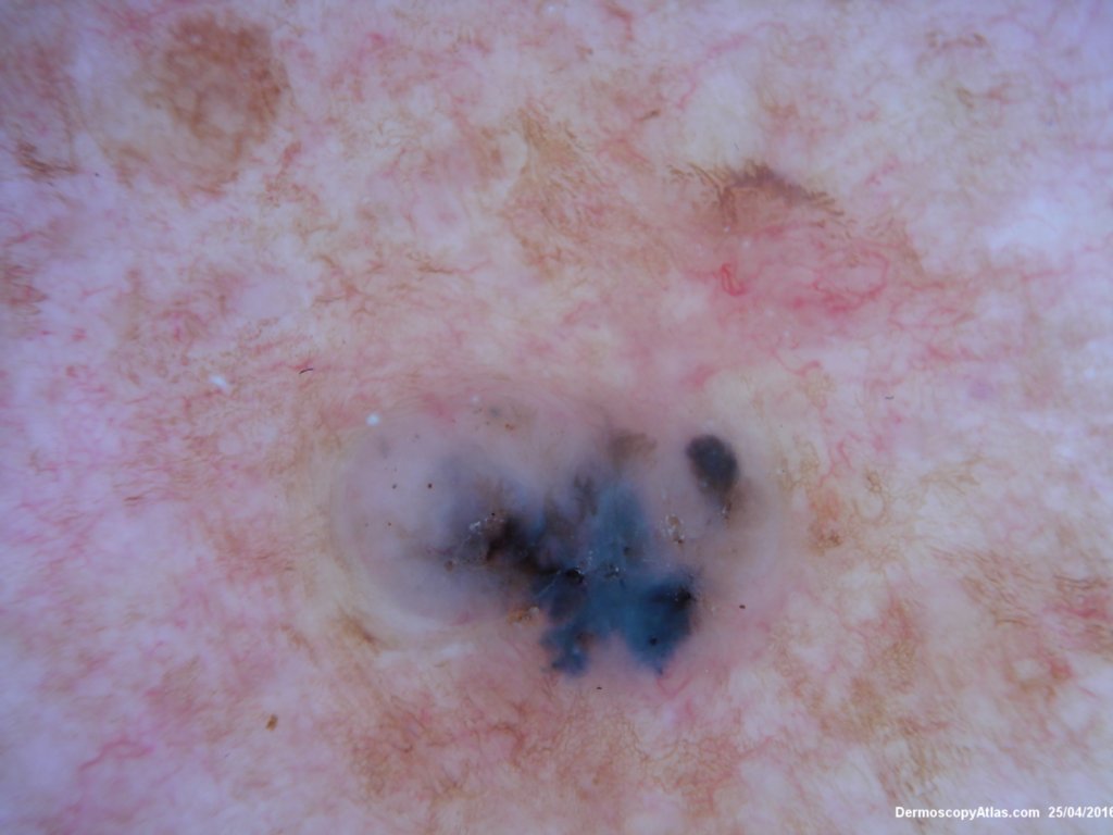
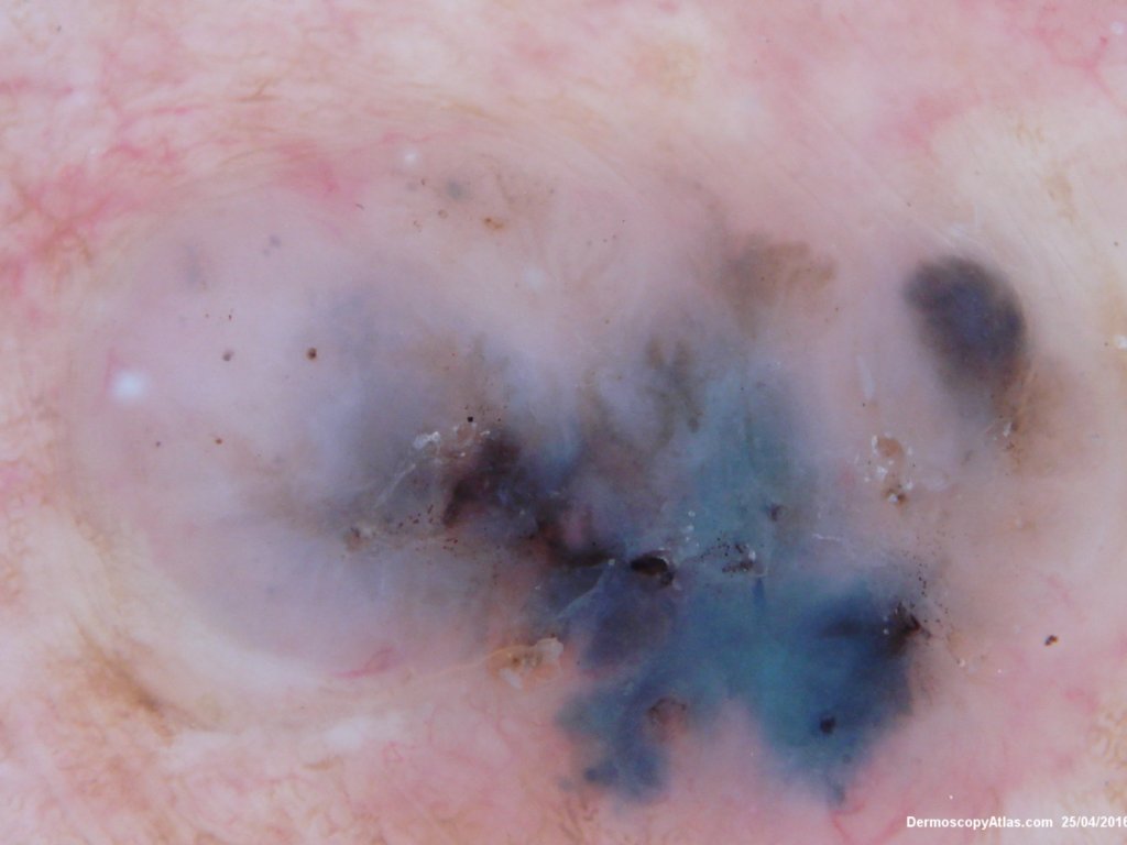
Description: Blue nodule on the upper back

History:

 Elderly man noted to have this pigmented lesion on his upper back during a skin check. Histology showed a pigmented BCC.

The dermatoscopy was made uip of blue and grey clods , some grey dots and pale structureless areas. There were also some lines radial radiating from a point suggestive of "spokewheels" and BCC.

DermNetNZ   eMedicine   PubMed   Dermatology Online   Archives   JAAD for "BCC pigmented"

  • Recent Submissions
  • Search
  • Diagnosis
  • Quiz
  • Image comparison
  • Submit an Image
  • Teaching Cases
  • Index
  • About

Copyright 2022. Dermoscopy Atlas. ALL Rights Reserved.

Change Image