Loading
TeleDermConsult-logo
  • Recent Submissions
  • Search
  • Diagnosis
  • Quiz
  • Image comparison
  • Submit
  • Teaching Cases
  • Index
  • About

<< Back to Diagnosis Page

Image Number #3392 (Melanoma invasive)

Site: Knee

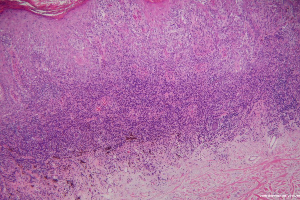
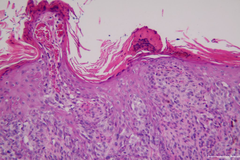
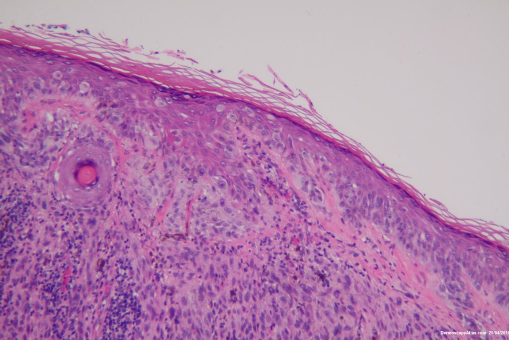
Diagnosis: Melanoma invasive

Sex: M

Age: 57

Type: Dermlite Polarised

Submitted By: Ian McColl

View Full Size Image

Click Here to View Details

Description: Lesion on the thigh

History:

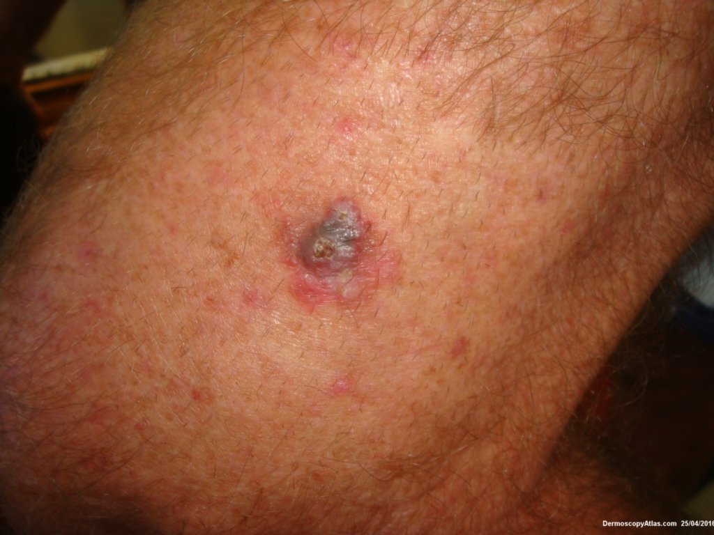
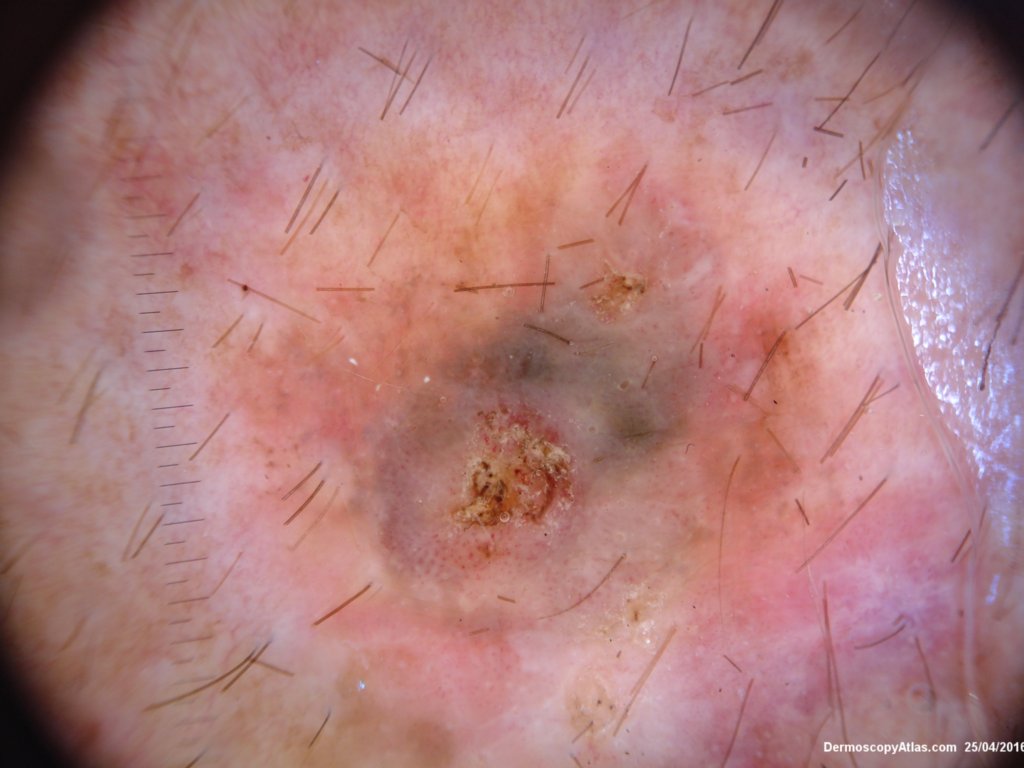
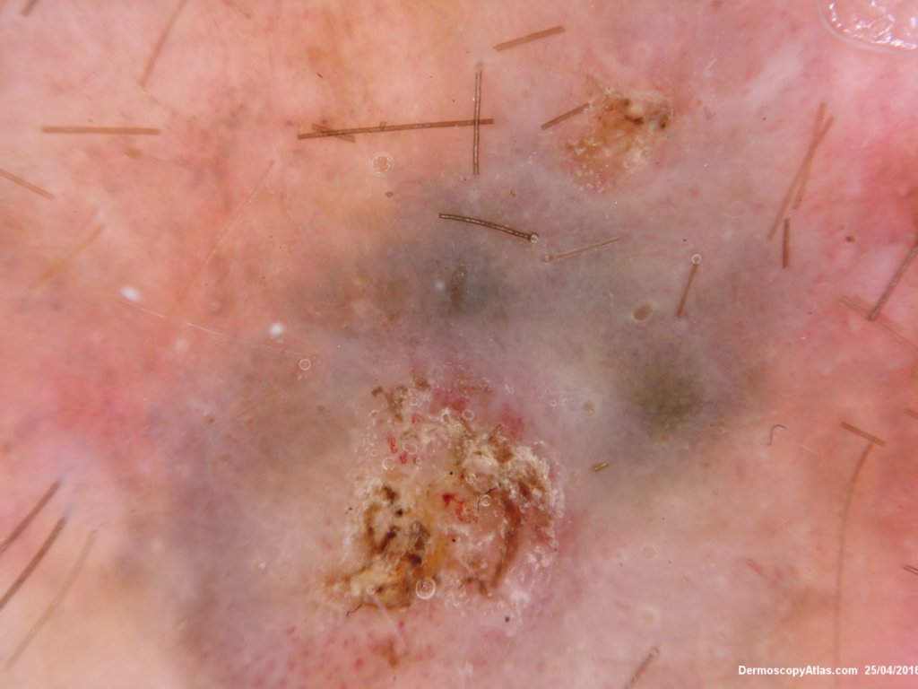
 This 57 years old male presented with a lesion on his thigh that he said had only been present for 6 weeks. It looked and felt keratotic and had a bluish discolouration associated with it. It was thought to be an irritated Seborrhoeic keratosis perhaps with some bleeding into it to account for the blue colour.


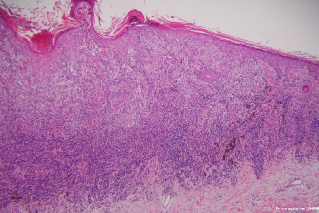
The shave biopsy was reported as a level 4 Invasive melanoma 1.7 mm thick with 3 mitoses per high powered field. There were no groin glands enlarged clinically.

DermNetNZ   eMedicine   PubMed   Dermatology Online   Archives   JAAD for "Melanoma invasive"

  • Recent Submissions
  • Search
  • Diagnosis
  • Quiz
  • Image comparison
  • Submit an Image
  • Teaching Cases
  • Index
  • About

Copyright 2022. Dermoscopy Atlas. ALL Rights Reserved.

Change Image