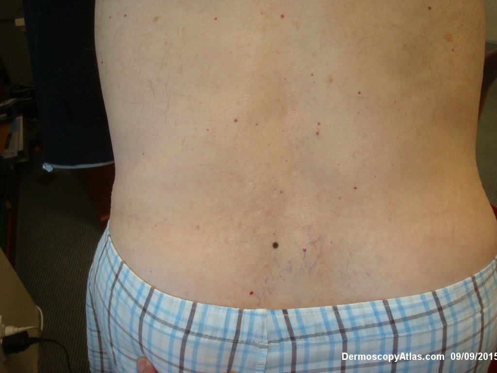
Loading
TeleDermConsult-logo
  • Recent Submissions
  • Search
  • Diagnosis
  • Quiz
  • Image comparison
  • Submit
  • Teaching Cases
  • Index
  • About

<< Back to Diagnosis Page

Image Number #3388 (Nevus junctional dysplastic lentiginous)

Site: Back

Diagnosis: Nevus junctional dysplastic lentiginous

Sex: M

Age: 92

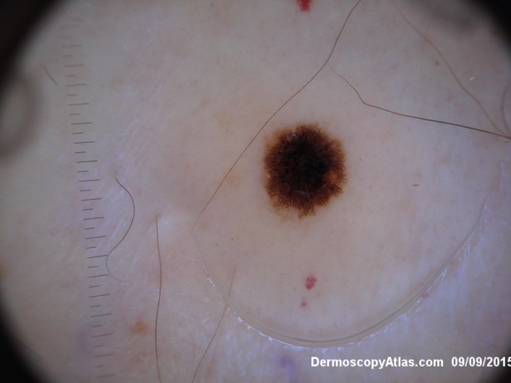
Type: Dermlite Polarised

Submitted By: Ian McColl

View Full Size Image

Click Here to View Details

Description: Dermatoscopy

History:

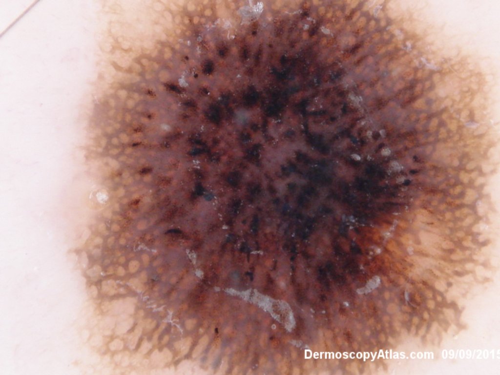
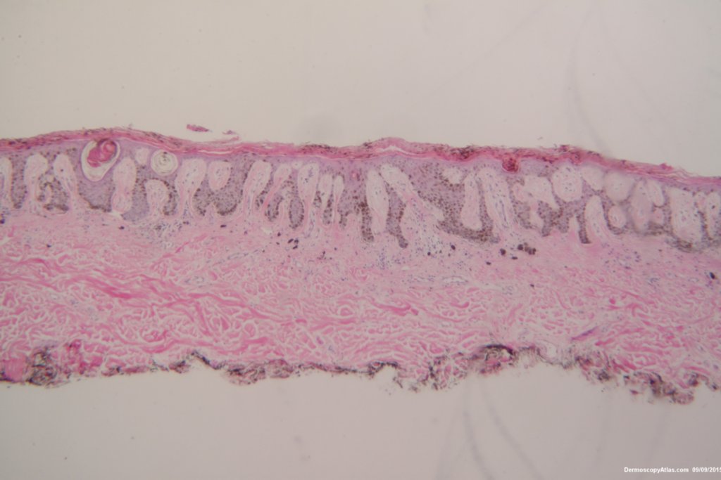
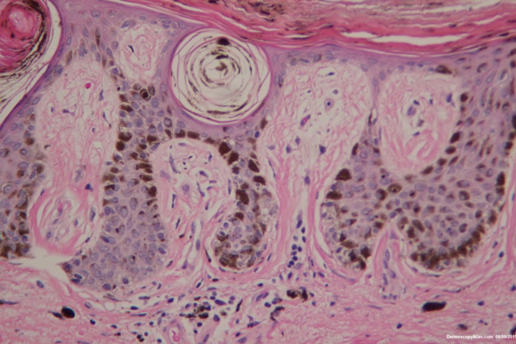
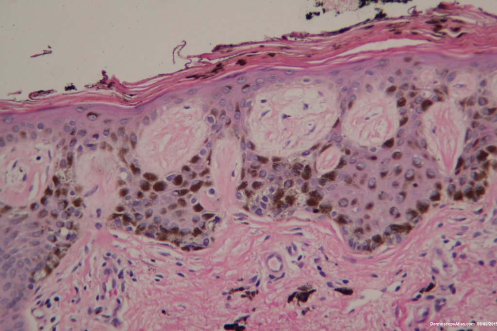
 Patient aged 92 noted to have this new lesion on the lower back. Note the extruded melanin on the stratum corneum and the lentiginous spread of melanocytes at the dermo epidermal junction. The dermatoscopy is symmetrical with multiple central clods presumably from the pigment in the stratum corneum. Tape stripping would have helped here. 

Reported as a benign dysplastic junctional nevus. Special stains failed to show any upward spread of the melanocytes.

View the video 

DermNetNZ   eMedicine   PubMed   Dermatology Online   Archives   JAAD for "Nevus junctional dysplastic lentiginous"

  • Recent Submissions
  • Search
  • Diagnosis
  • Quiz
  • Image comparison
  • Submit an Image
  • Teaching Cases
  • Index
  • About

Copyright 2022. Dermoscopy Atlas. ALL Rights Reserved.

Change Image