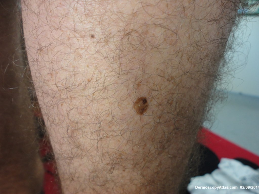
Loading
TeleDermConsult-logo
  • Recent Submissions
  • Search
  • Diagnosis
  • Quiz
  • Image comparison
  • Submit
  • Teaching Cases
  • Index
  • About

<< Back to Diagnosis Page

Image Number #3362 (Seborrhoeic keratosis)

Site: Calf

Diagnosis: Seborrhoeic keratosis

Sex: M

Age: 51

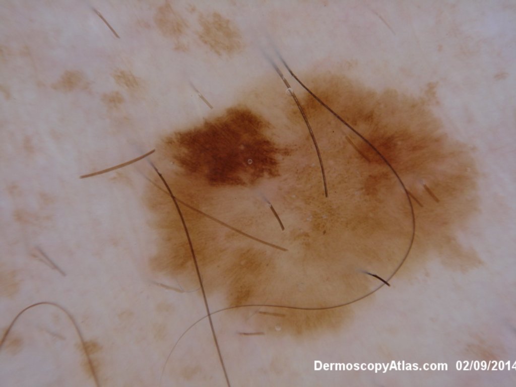
Type: Dermlite Polarised

Submitted By: Ian McColl

View Full Size Image

Click Here to View Details

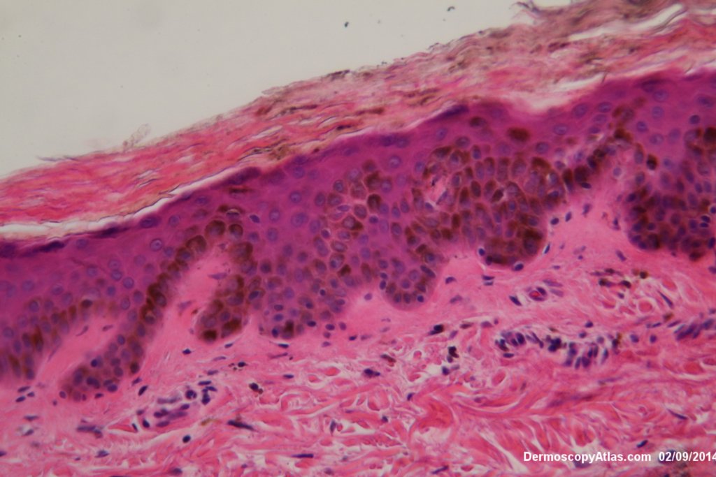
Description: Hyperkeratosis with acanthosis and hyperpigmentation No increase in melanocytes

History:

 This man presented for a skin check. He was noted to have a lesion on his calf with eccentric hyperpigmentation which on high power view showed some dots as lines and a structureless picture elsewhere. Pigmented IEC was suspected but it was a seborrhoeic keratosis on histology.

See the initial part of this video for a discussion of the histology.

DermNetNZ   eMedicine   PubMed   Dermatology Online   Archives   JAAD for "Seborrhoeic keratosis"

  • Recent Submissions
  • Search
  • Diagnosis
  • Quiz
  • Image comparison
  • Submit an Image
  • Teaching Cases
  • Index
  • About

Copyright 2022. Dermoscopy Atlas. ALL Rights Reserved.

Change Image