
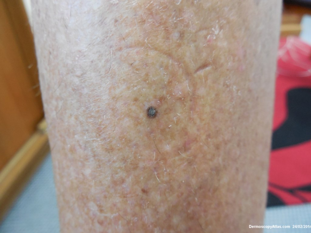
Site: Leg
Diagnosis: Seborrhoeic keratosis
Sex: F
Age: 61
Type: Dermlite Polarised
Submitted By: Ian McColl
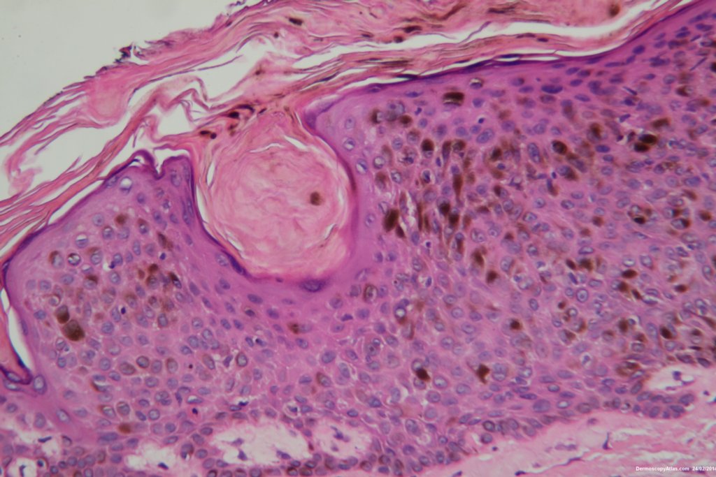
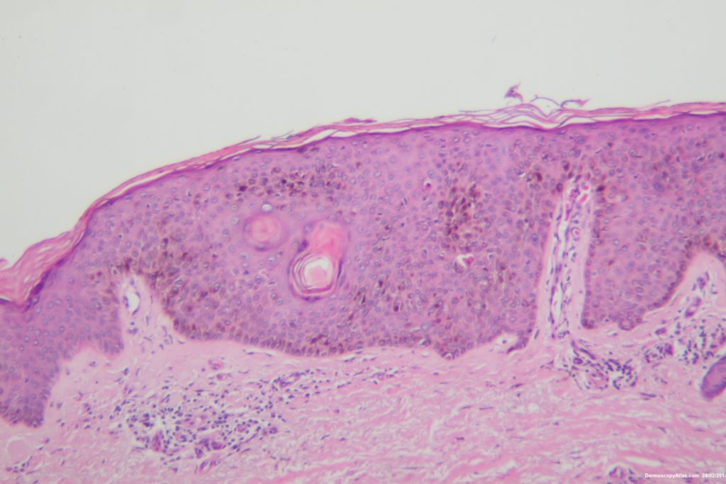
Description: Histology Pigmented seborrhoeic keratosis
History:
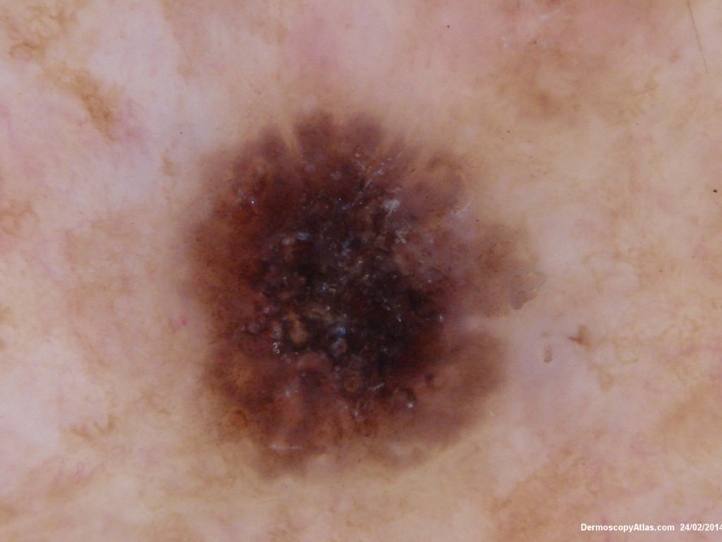
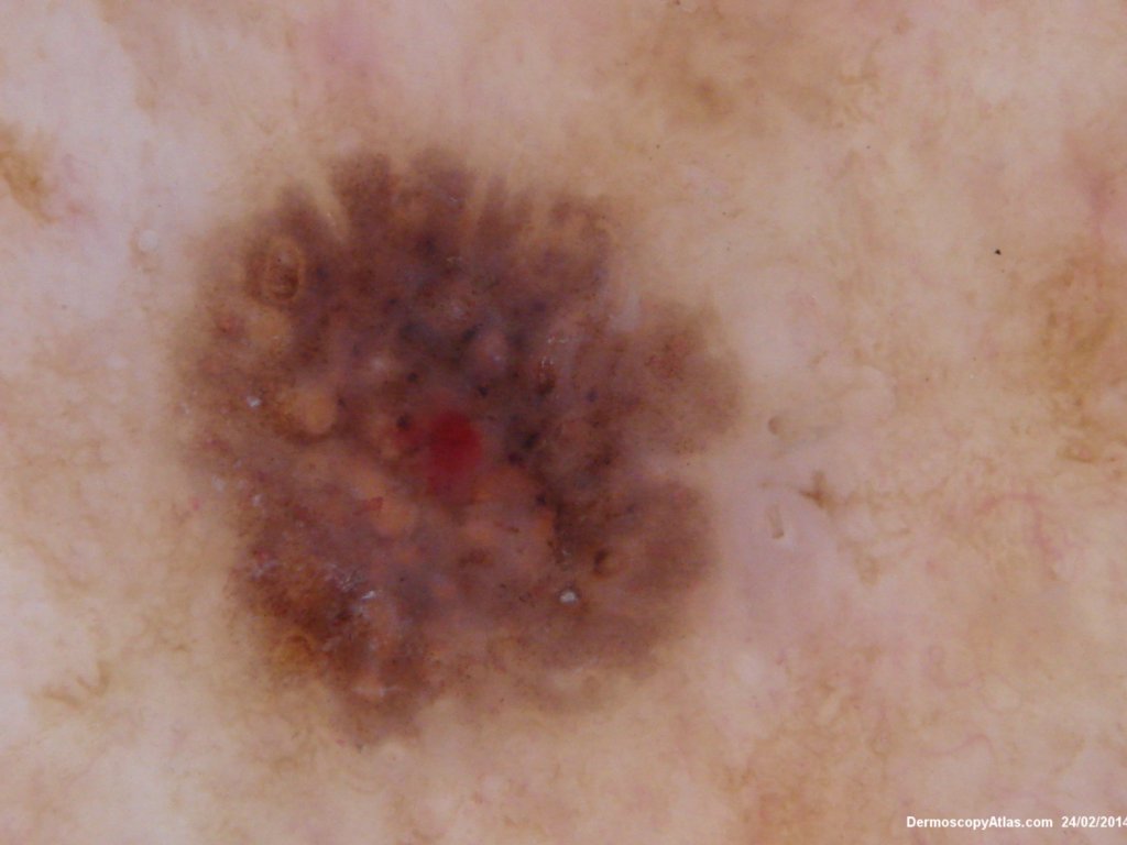
This lesion had been present on the lower leg anteriorly for 2 years. Slowly becoming larger with a densely adherent surface scale. The scale was removed to reveal the underlying dermatoscopic features with different coloured clods and no real network.
The histology shows a heavily pigmented seborrhoeic keratosis with pigment cells being seen in the stratum corneum explaining the black colour.