
Site: Leg
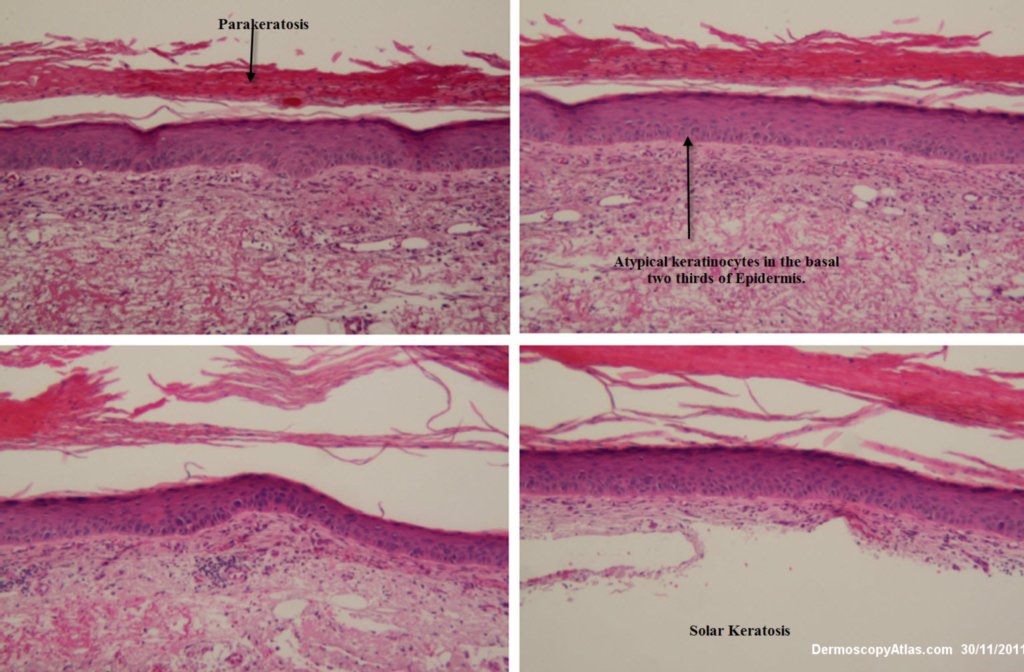
Diagnosis: Pigmented solar keratosis
Sex: M
Age: 57
Type: Heine
Submitted By: Ian McColl
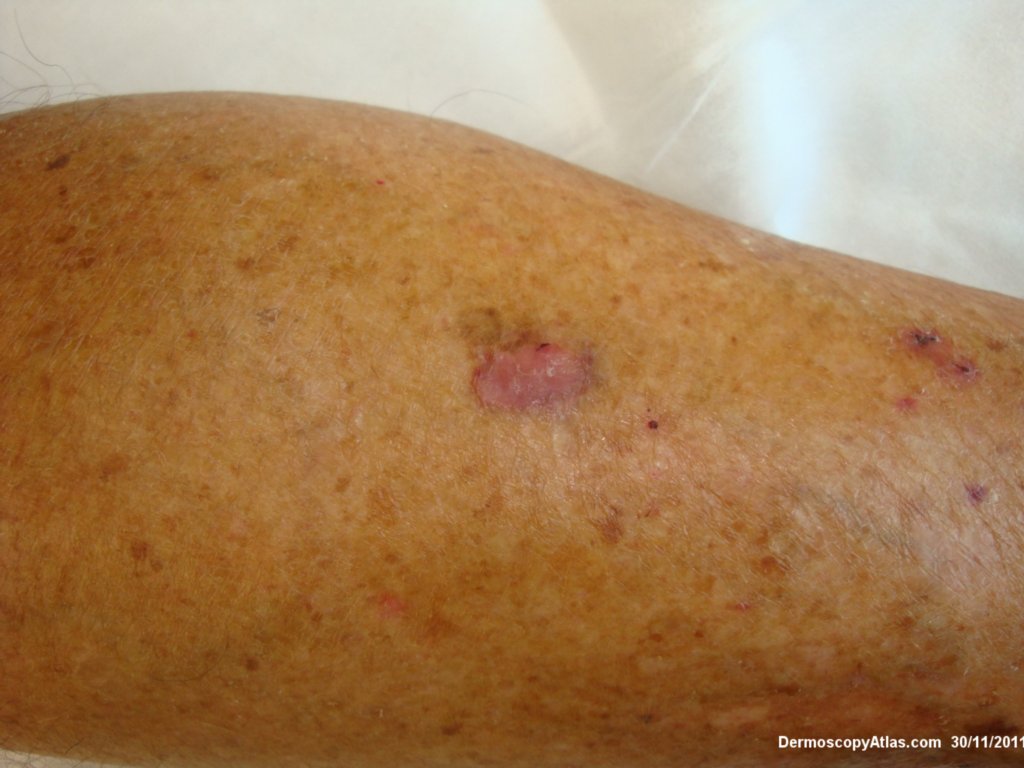
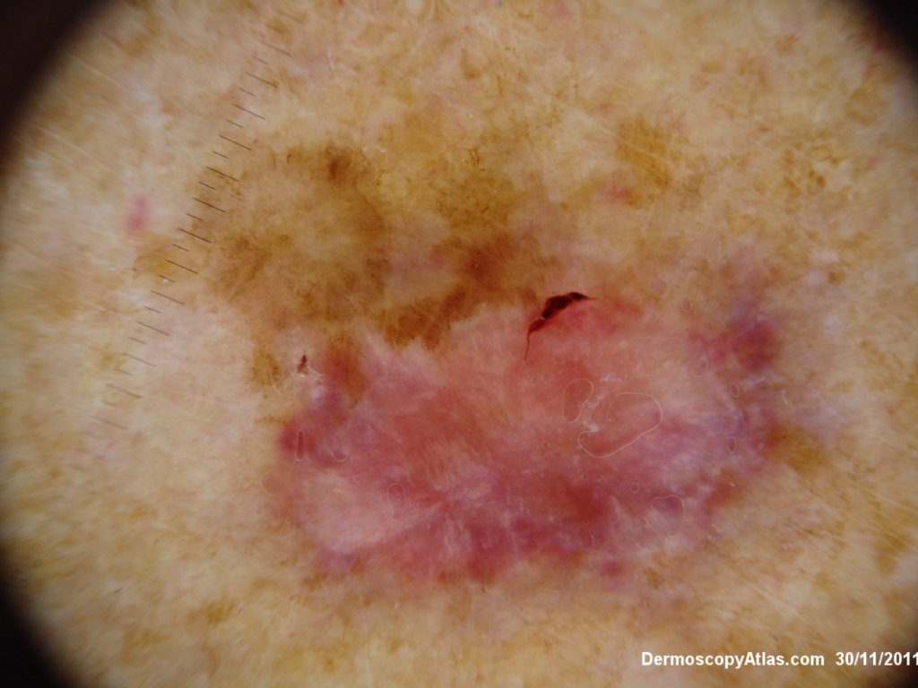
Description: Lesion on calf with pink and surrounding pigmented area
History:
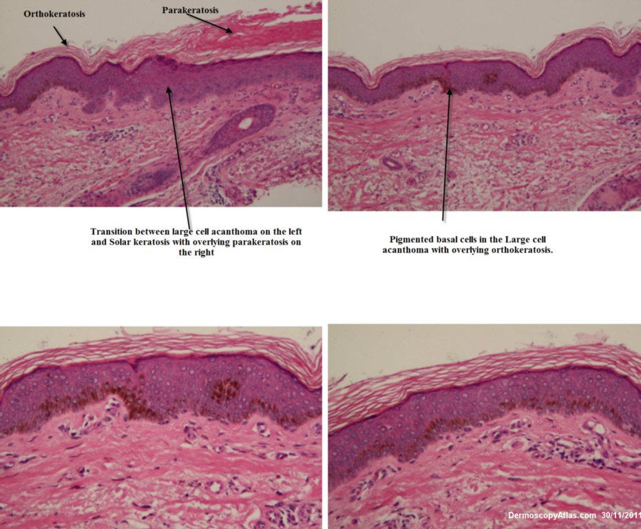
This lesion was on a man's calf. The pink area was adjacent to a pigmented area. This combination of pink and brown always makes you consider melanoma especially with the white lines in the pink area. Clinically the edge of the pink area might also suggest a porokeratosis. A large shave to include part of the pink and the brown areas revealed the pink area to be a solar keratosis and the pigmented area to be a large cell acanthoma. The latter is probably a seb k variant with keratinocytes about double the normal size and larger nucleii. Atypia though is unusual and there is orthokeratosis of the overlying stratum corneum rather than parakeratosis.