
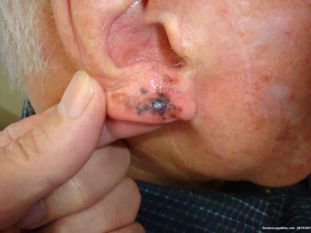
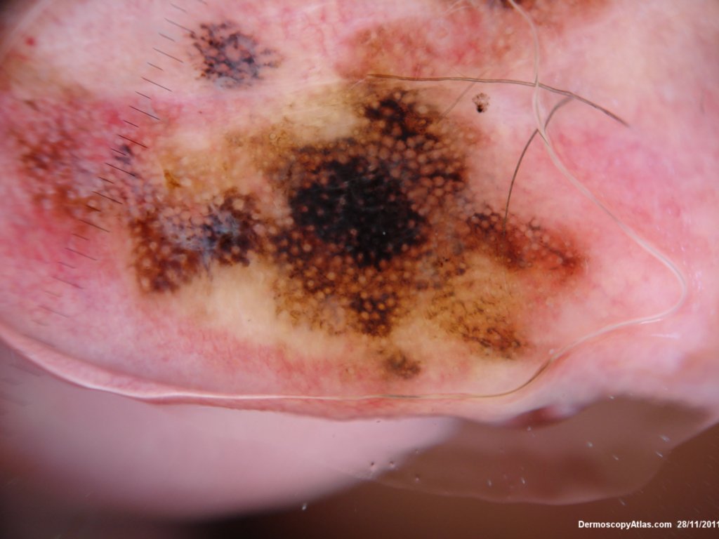
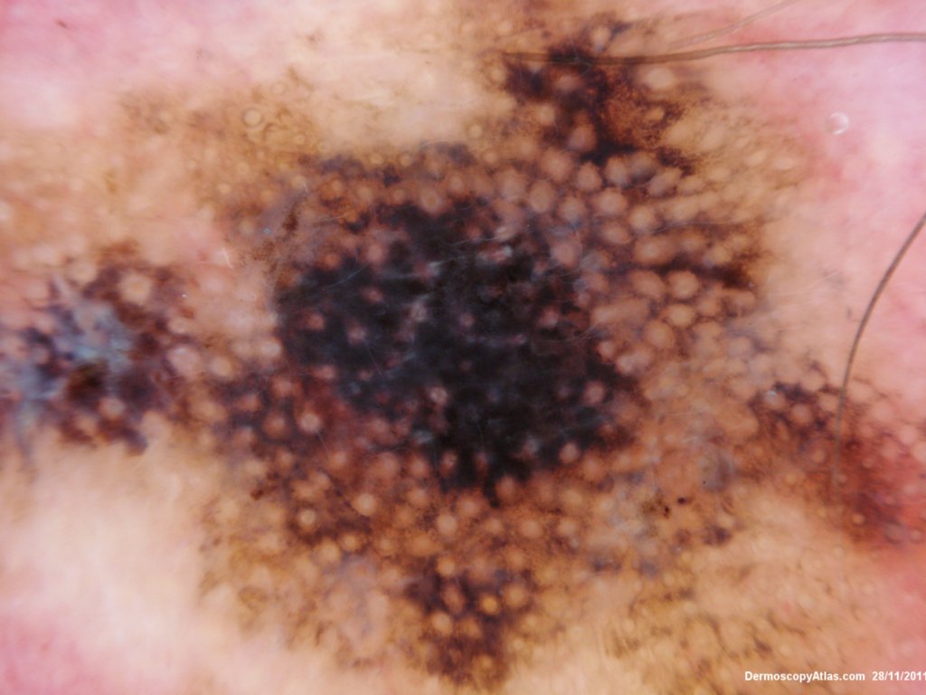
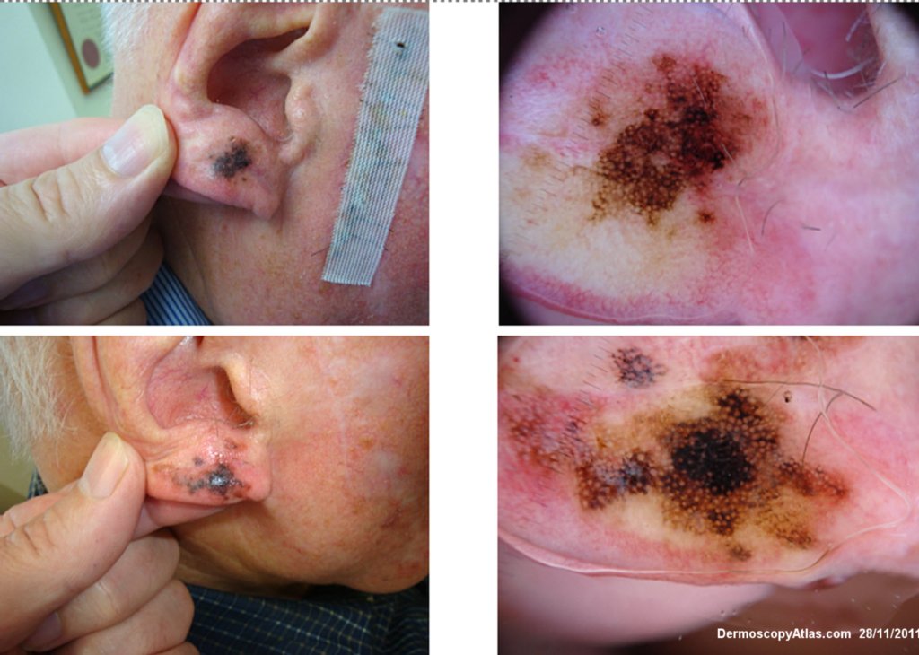
Site: Ear lobe
Diagnosis: Lentigo Maligna
Sex: M
Age: 86
Type: Dermlite Polarised
Submitted By: Ian McColl
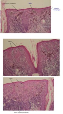
Description: Pigmented lesion on the ear
History:
This elderly male had a pigmented lesion on his ear which was shown to be a lentigo maligna but surgery was refused. It progressed over 5 years as shown but remained a lentigo maligna or melanoma in situ. There is marked perifollicular pigmentation with black structureless areas where the melanoma has obliterated the follicles. Sometimes the terms annular granular structures and rhomboid figures are used along with asymmetrical perifollicular pigmentation to describe these earlier changes. In truth grey circles are often the earliest features of lentigo maligna particuarly on the face.