
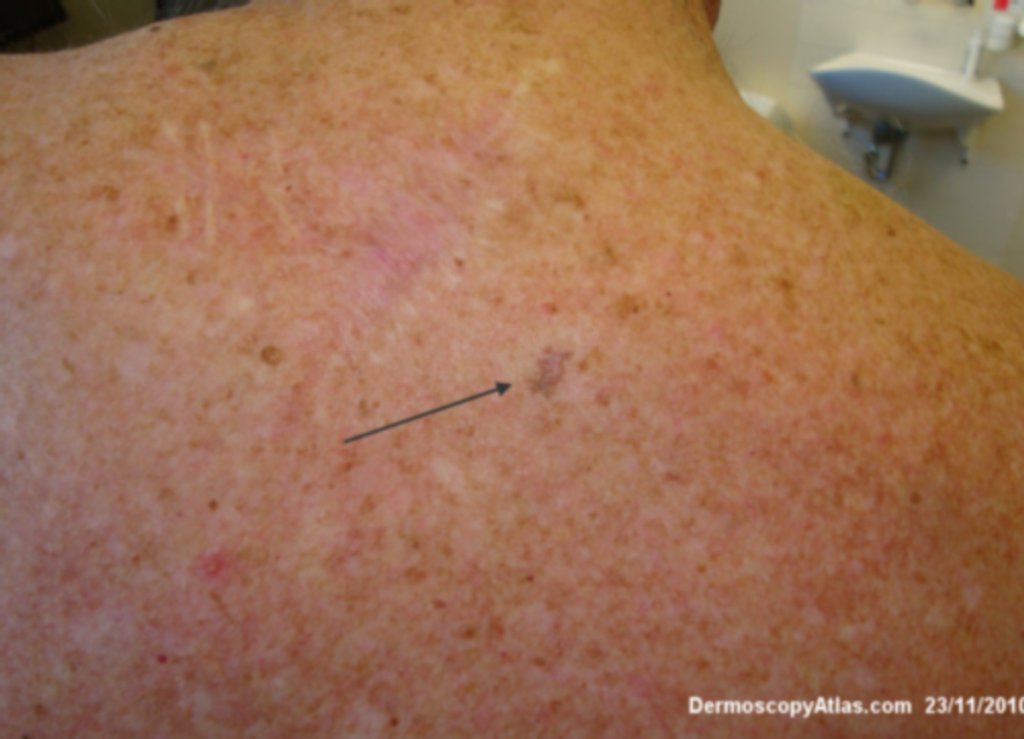
Site: Back
Diagnosis: Melanoma in situ
Sex: M
Age: 55
Type: Dermlite Non Polarised
Submitted By: Ian McColl
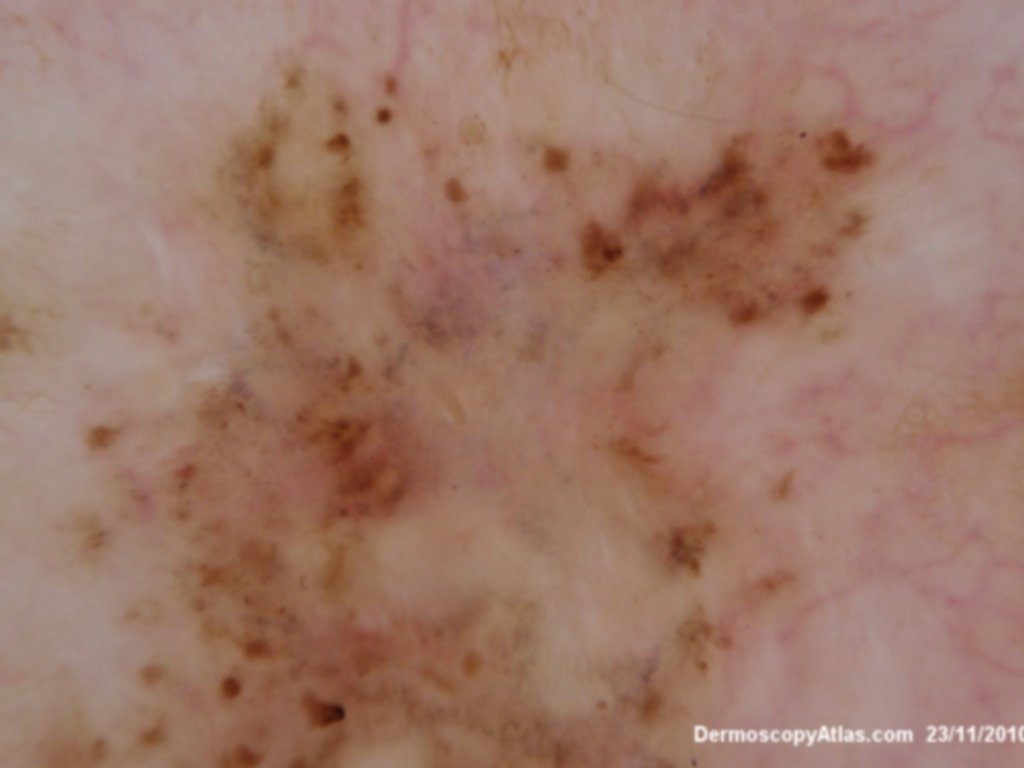
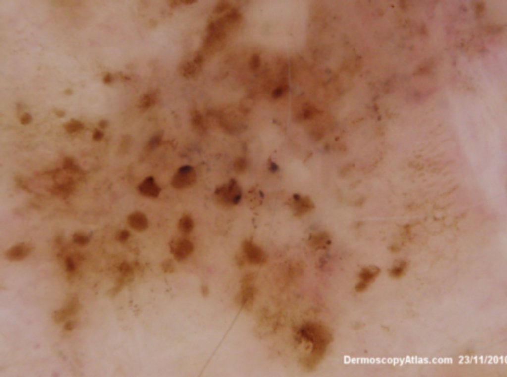
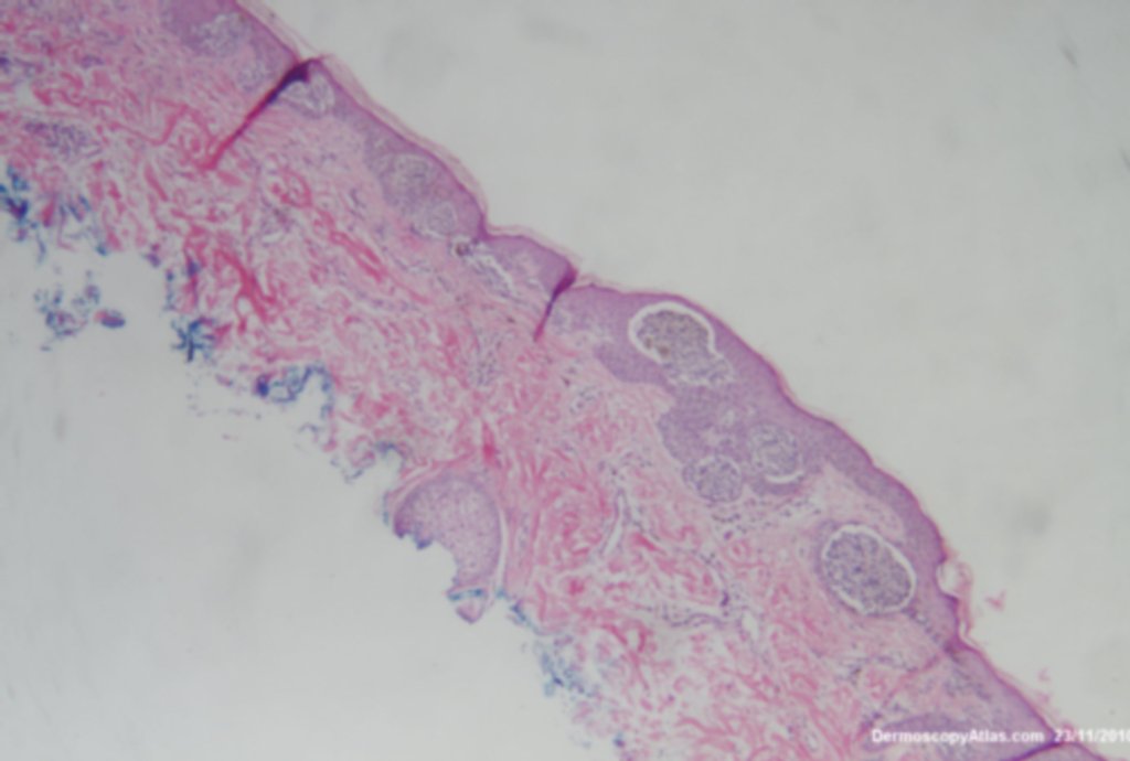
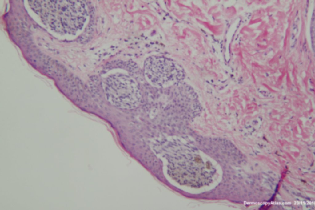
Description: Close up histology Nests of atypical melanocytes Melanoma in situ with markedly atypical junctional melanocytic proliferation including a focal lentiginous pattern and regressional changes. No Pagetoid spread. Pigmentary incontinence in the upper dermis. (Hence the grey dots)
History:
A man in his late 50s had this lesion noted when a full skin check was being performed. It was a melanoma in situ with markedly atypical junctional melanocytic proliferation including a focal lentiginous pattern and regressional changes. No Pagetoid spread. Pigmentary incontinence in the upper dermis. (Hence the grey dots) The brown and sometimes black clods represent nests of melanocytes at the dermoepidermal junction , sometimes pushing higher into the epidermis giving the darker colour. The size of the clods is due to the varying size of the nests.