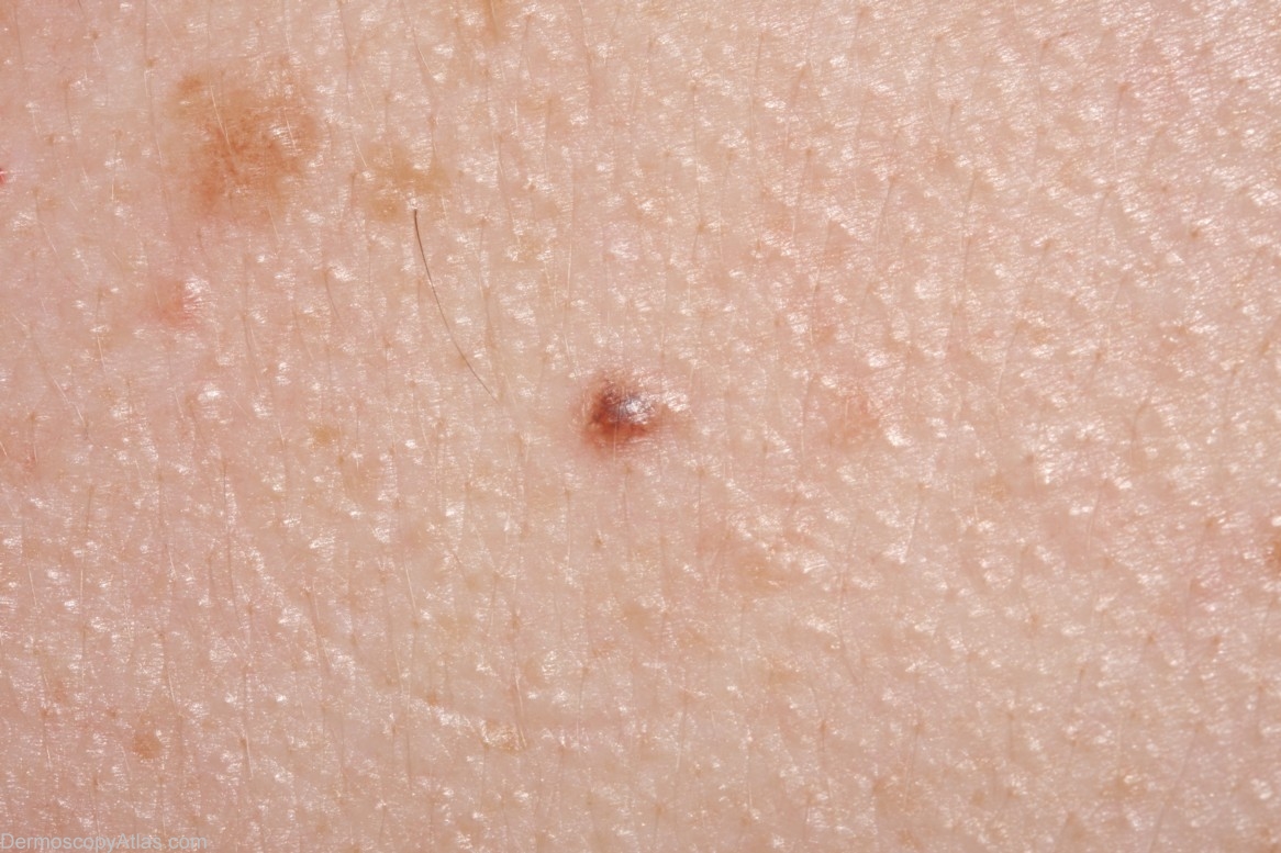
Loading
TeleDermConsult-logo
  • Recent Submissions
  • Search
  • Diagnosis
  • Quiz
  • Image comparison
  • Submit
  • Teaching Cases
  • Index
  • About

<< Back to Diagnosis Page

Image Number #3024 (Nevus junctional)

Site: Back

Diagnosis: Nevus junctional

Sex: M

Age: 20

Type: Dermlite Non Polarised

Submitted By: Cliff Rosendahl

View Full Size Image

Click Here to View Details

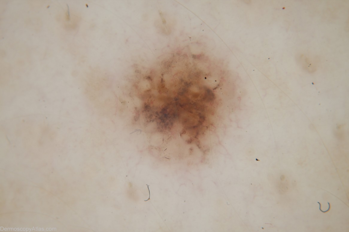
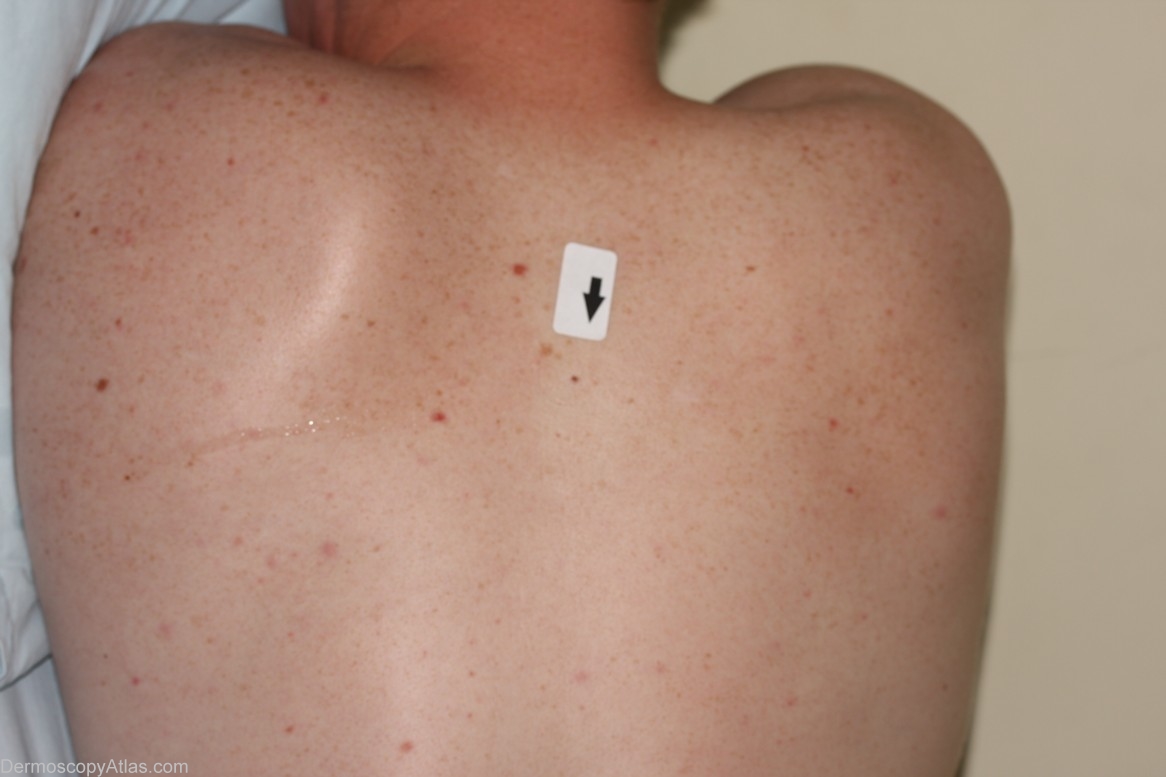
Description: Macro image

History:

This 20 year old male has a strong family history of melaoma - His father has had 3 level 1 melanomas by age 44. This was excised and the histology report read "...junctional naevus with mild to moderate atypia. Advanced lymphocytic regression with melanin incontinence and heavy lymphocytic infiltrate. So much regression is present that I prefer to call this atypical rather than dysplastic."

DermNetNZ   eMedicine   PubMed   Dermatology Online   Archives   JAAD for "Nevus junctional"

  • Recent Submissions
  • Search
  • Diagnosis
  • Quiz
  • Image comparison
  • Submit an Image
  • Teaching Cases
  • Index
  • About

Copyright 2022. Dermoscopy Atlas. ALL Rights Reserved.

Change Image