
Site: Temple
Diagnosis: Lentigo Maligna
Sex: M
Age: 80
Type: Heine
Submitted By: Jean-Yves Gourhant

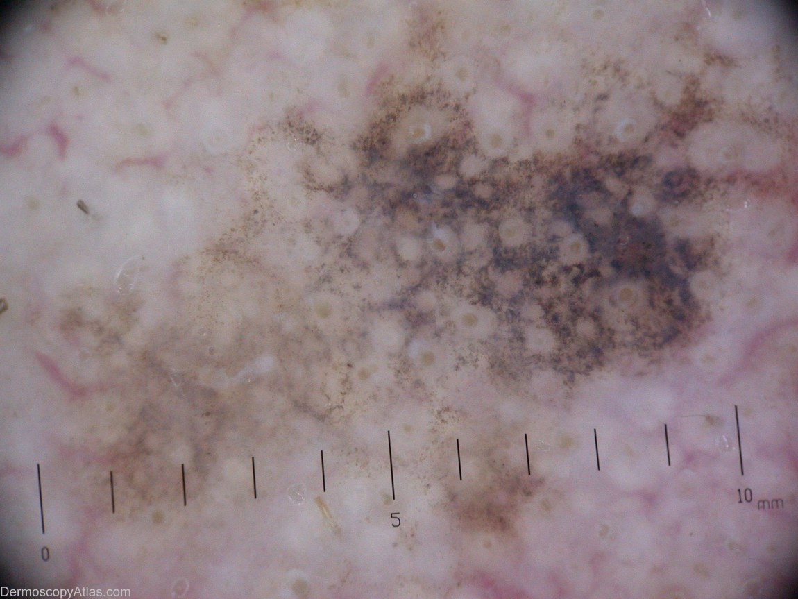
Description: Dermoscopy: asymetrically pigmented follicular openings (APFO), grey dots forming a granular pseudo-network, rhomboidal structures by place, obliterated hair follicles.
History: This 79 years gentleman consulted for actinic keratoses on his hands. This dark spot on the temple was seen during the exam. The dermoscopic exam, as pointed out by Alex Chamberlain, shows the evolution of a Lentigo Maligna: asymetric pigmentation around the follicular openings, grey dots forming an annular-granular pseudo network, which intensifies into rhomboidal structures; the pigmentation hides the follicles. The pathology confirmed the provisional diagnosis of Lentigo Maligna (Melanoma in situ).