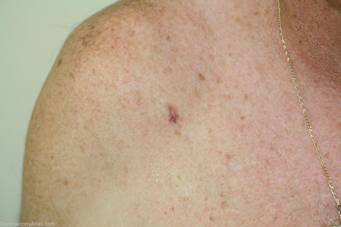
Loading
TeleDermConsult-logo
  • Recent Submissions
  • Search
  • Diagnosis
  • Quiz
  • Image comparison
  • Submit
  • Teaching Cases
  • Index
  • About

<< Back to Diagnosis Page

Image Number #2999 (Pigmented basal cell carcinoma)

Site: Shoulder

Diagnosis: Pigmented basal cell carcinoma

Sex: M

Age: 66

Type: Dermlite Non Polarised

Submitted By: Cliff Rosendahl

View Full Size Image

Click Here to View Details

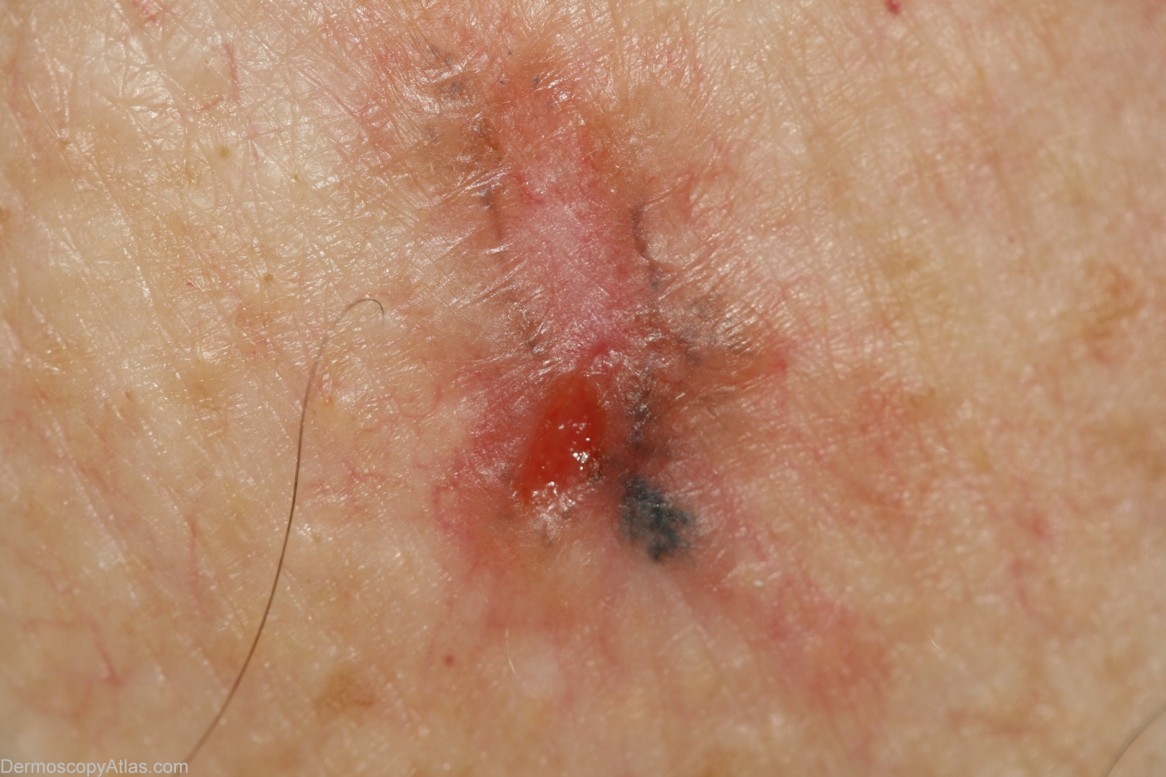
Description: dermoscopic image. This BCC has branched (arborising) vessels,focal peripheral lines radial (spoke-wheel structures), ulceration, vivid blue clods, scarring, and "dishwater bown" areas

History: This 66 year old gentleman presented for a routine skin check. This lesion had not been noted 12 months previously. Histology was reported as a " solid and infiltrative pigmented BCC"

DermNetNZ   eMedicine   PubMed   Dermatology Online   Archives   JAAD for "Pigmented basal cell carcinoma"

  • Recent Submissions
  • Search
  • Diagnosis
  • Quiz
  • Image comparison
  • Submit an Image
  • Teaching Cases
  • Index
  • About

Copyright 2022. Dermoscopy Atlas. ALL Rights Reserved.

Change Image