
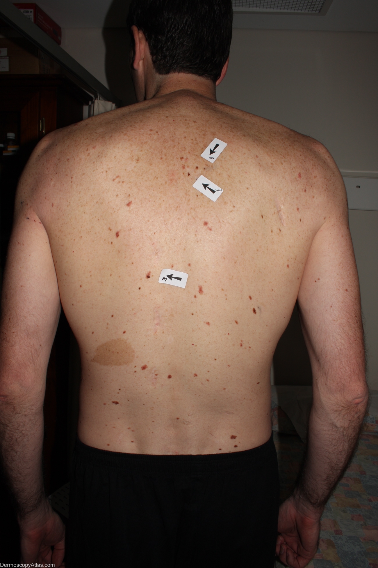
Site: Back
Diagnosis: Melanoma in situ
Sex: M
Age: 35
Type: Dermlite Non Polarised
Submitted By: Alan Cameron
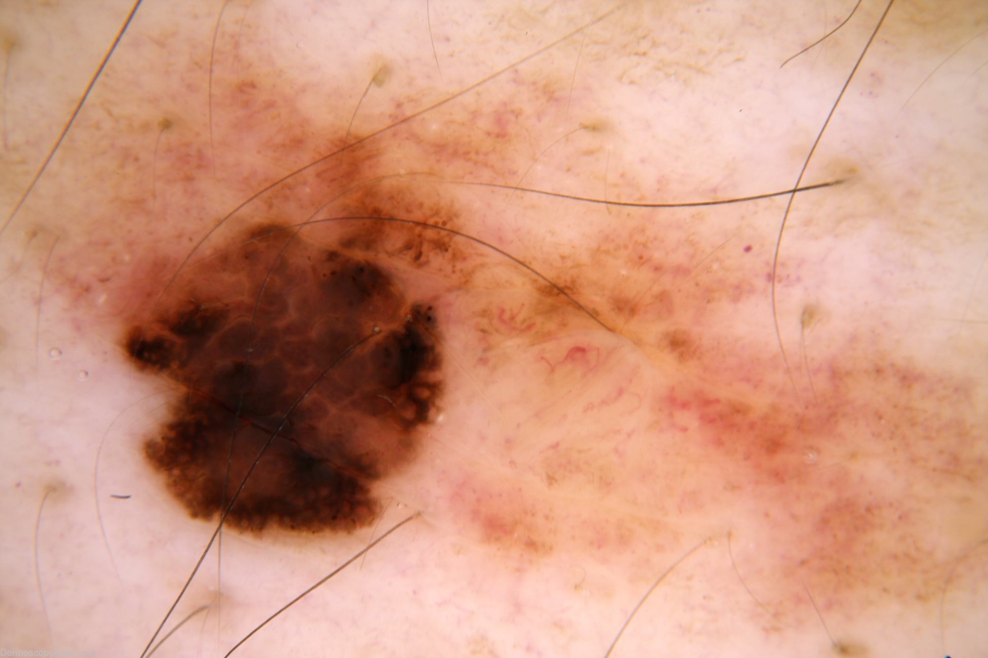
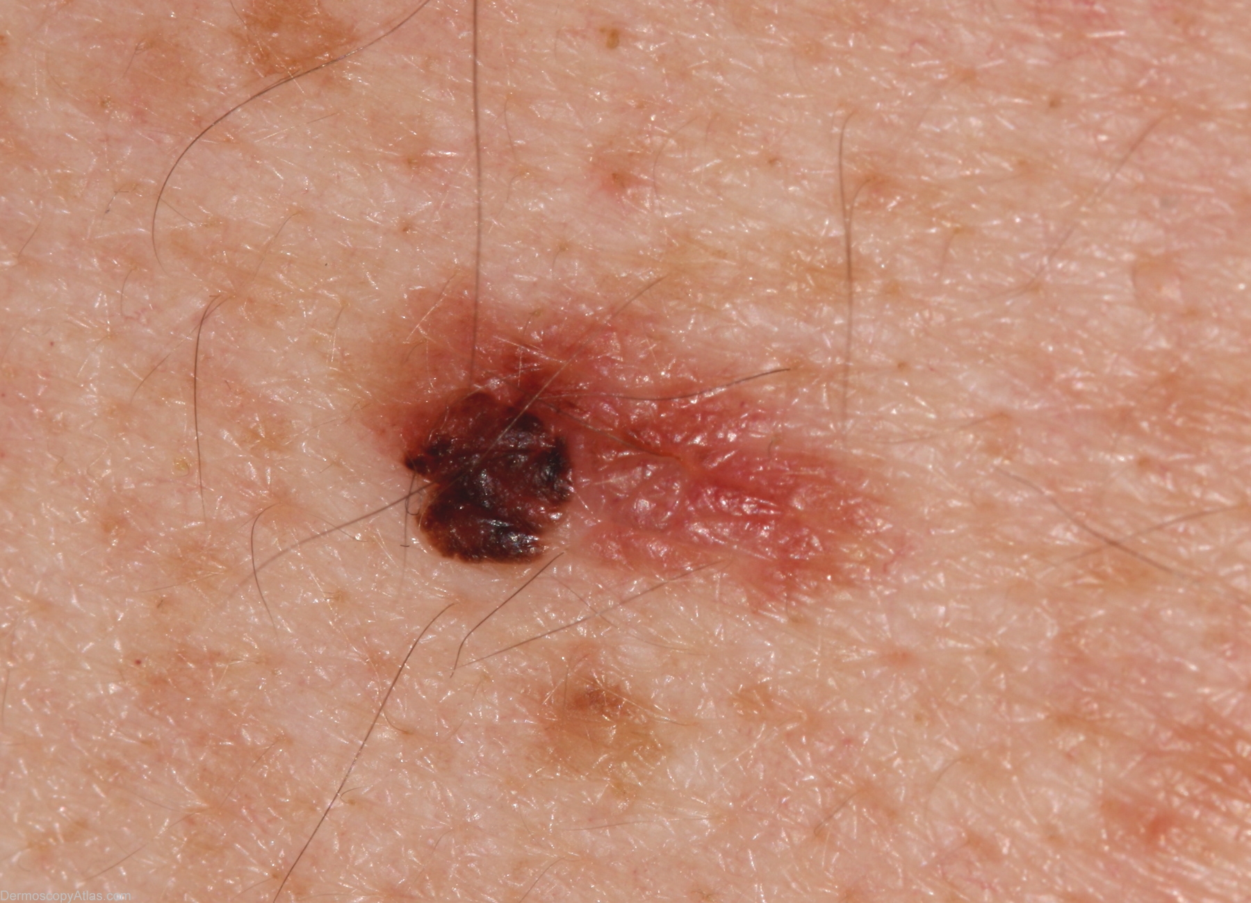
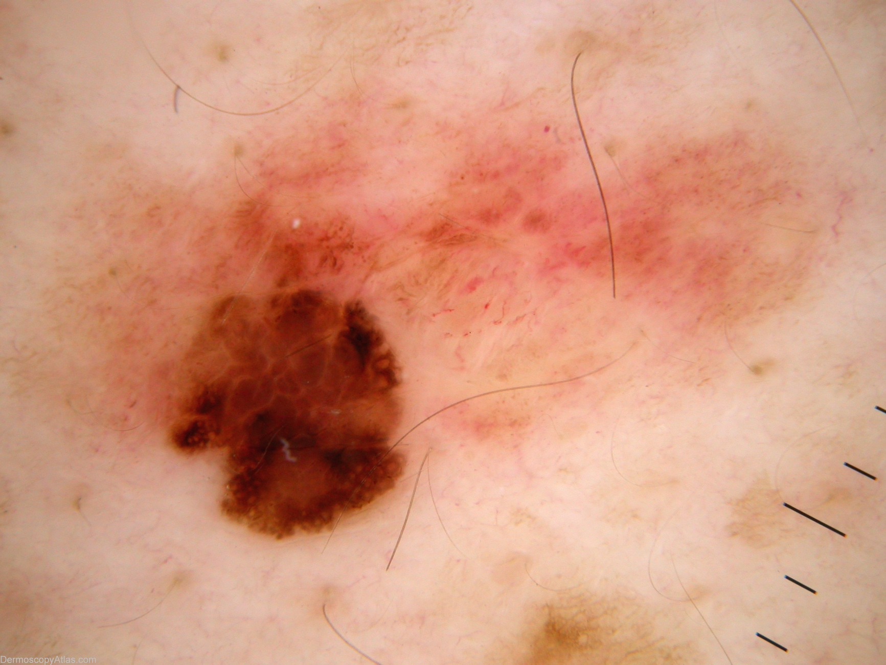
Description: Multiple atypical naevi. Melanoma is inferior lesion "M"
History: 35 year old male with atypical mole syndrome but no past history. Referred by GP regarding this lesion. Melanoma 394 was also found at this examination. Histology reported as; Sections show a level 1 (in situ) superficial spreading melanoma arising in a dysplastic compound naevus. There is no ulceration, dermal invasion, lymphocytic infiltrate or regression.