
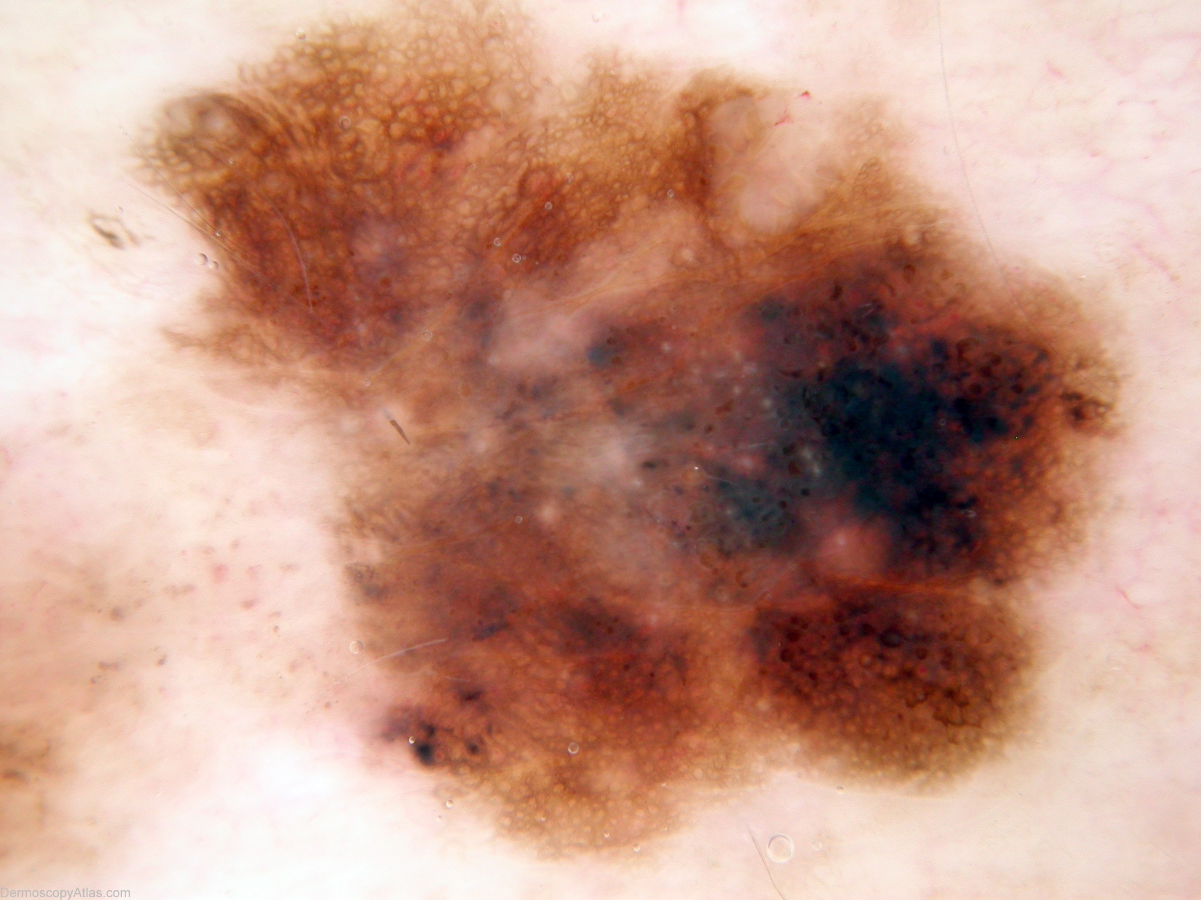
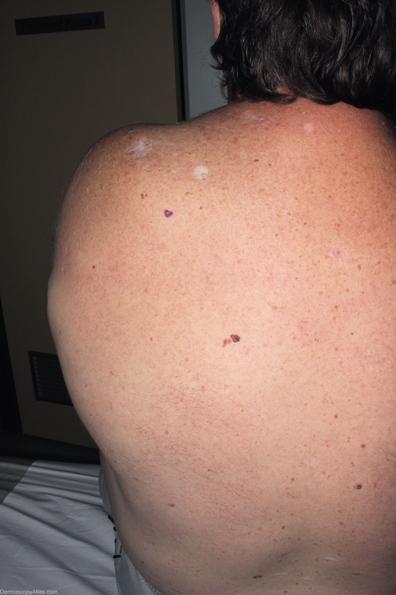
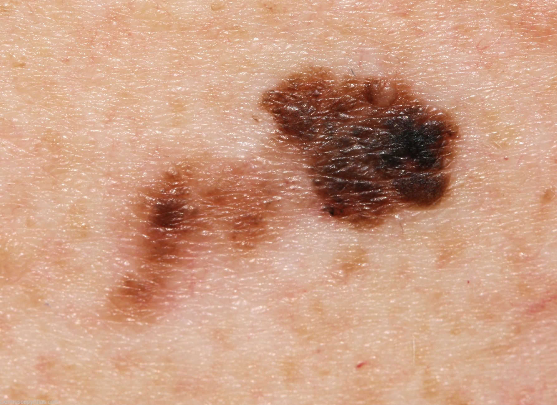
Site: Back
Diagnosis: Melanoma invasive
Sex: M
Age: 49
Type: Mixed
Submitted By: Alan Cameron
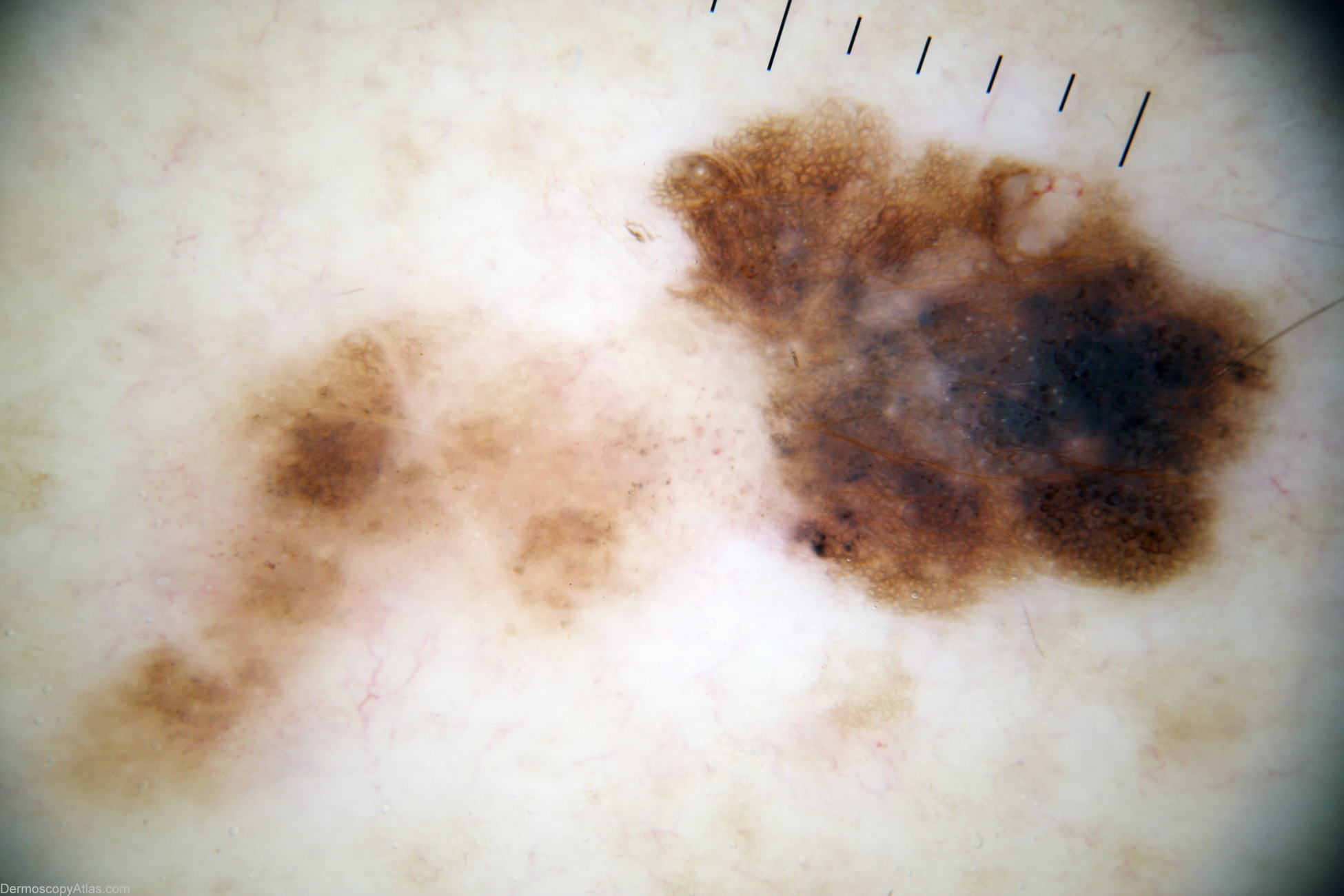
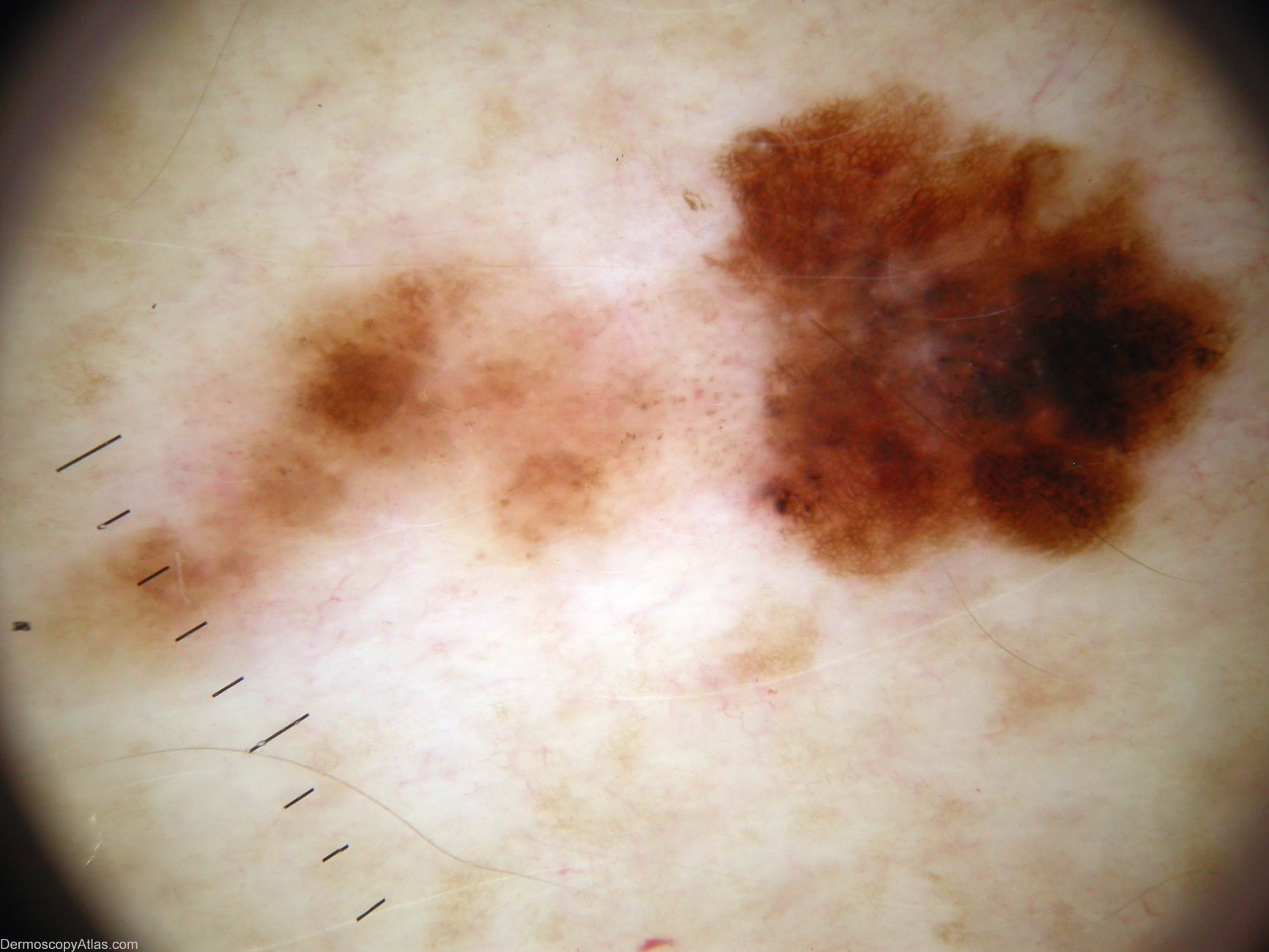
Description: Detail Fluid view highlighting white clods and orange/brown clods mimicking comedo-like openings. (detail lost due to file compression in previous dermoscopic views, apparent in originals)
History: 49 year old man with past history of several BCCs, not seen for 3 years. New lesion on back, patient not aware of. Histology reported as; Sections show regressing level 2 superficial spreading malignant melanoma arising in dysplastic naevus, and characterised by pigmented atypical melanocytes infiltrating epidermis in a pagetoid pattern, and focally into the papillary dermis in association with signs of regression. There are no mitoses or ulceration. TUMOUR THICKNESS 0.63MM