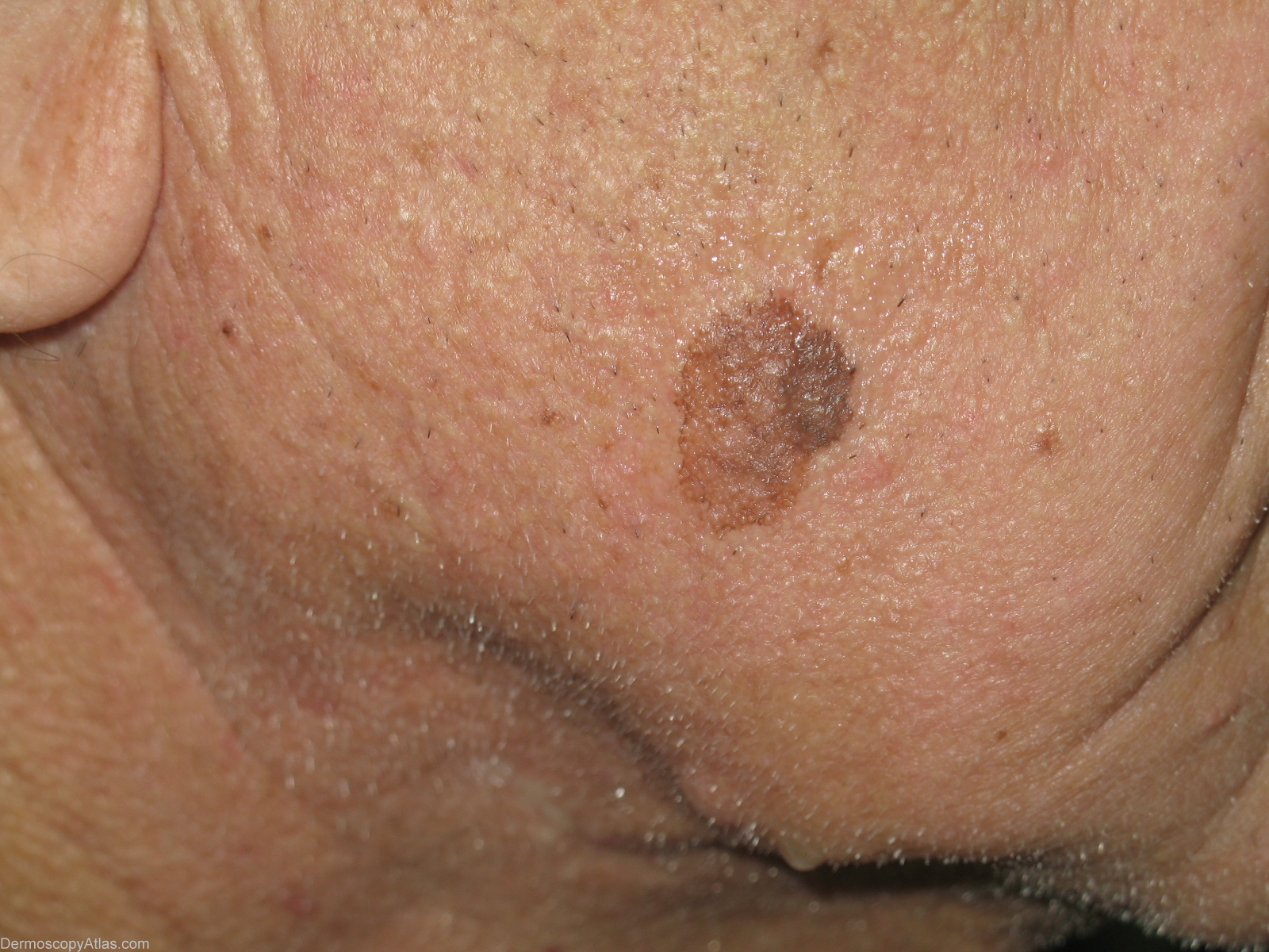
Loading
TeleDermConsult-logo
  • Recent Submissions
  • Search
  • Diagnosis
  • Quiz
  • Image comparison
  • Submit
  • Teaching Cases
  • Index
  • About

<< Back to Diagnosis Page

Image Number #2873 (Solar lentigo)

Site: Cheek

Diagnosis: Solar lentigo

Sex: M

Age: 83

Type: Heine

Submitted By: Stelios Minas

View Full Size Image

Click Here to View Details

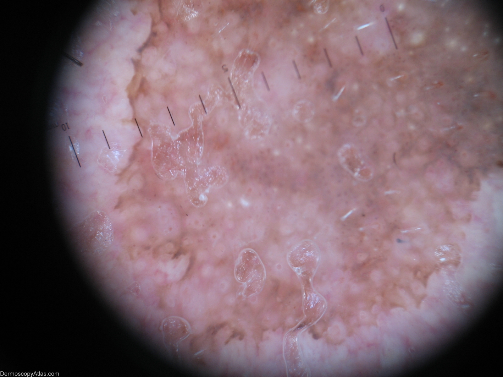
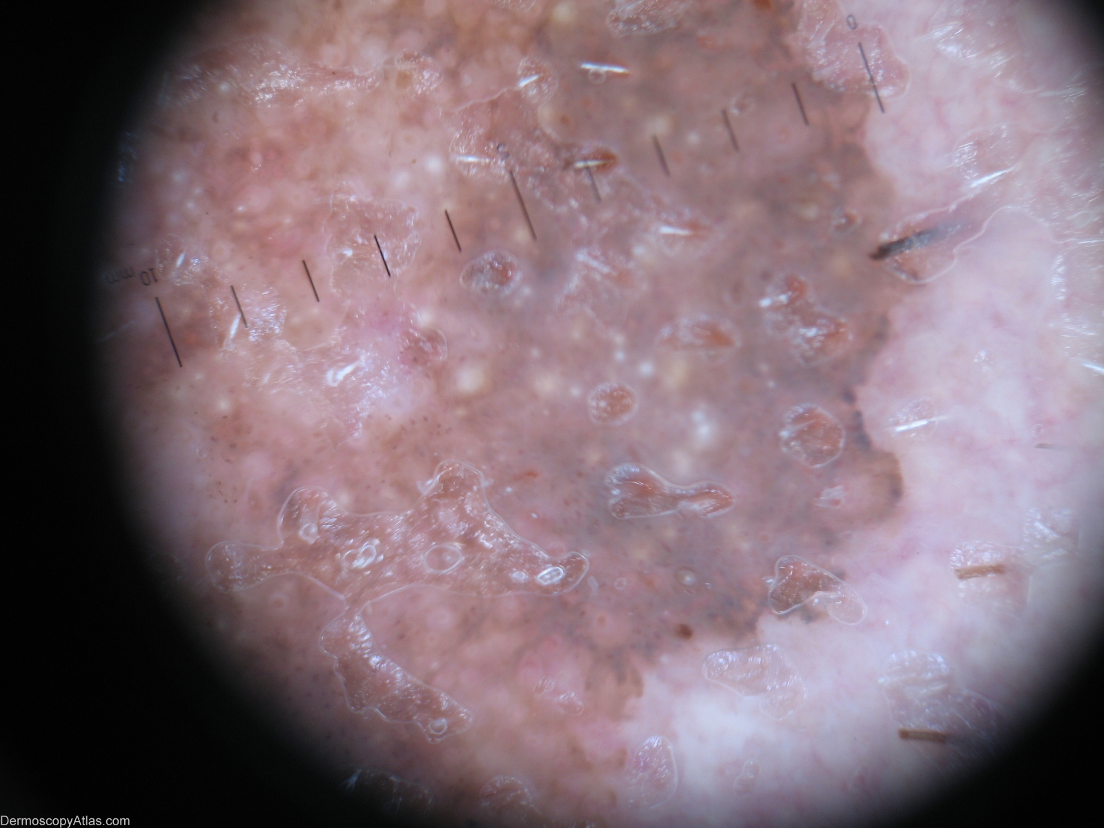
Description: Milia-like cysts and comedo-like openings.

History: Lentigo solar(left part) crossing over with a seborrhoeic keratosis(right part). A 83-year-old man developed an enlarging pigmented patch of his cheek.A biopsy was performed and pathology confirmed the diagnosis of solar lentigo. Dermoscopic description: Right part(seborrhoeic keratosis)- Milia-like cysts,comedo-like openings. Left part(lentigo solar)-sharply demarcated pigmented network and moth-eaten border.

DermNetNZ   eMedicine   PubMed   Dermatology Online   Archives   JAAD for "Solar lentigo"

  • Recent Submissions
  • Search
  • Diagnosis
  • Quiz
  • Image comparison
  • Submit an Image
  • Teaching Cases
  • Index
  • About

Copyright 2022. Dermoscopy Atlas. ALL Rights Reserved.

Change Image