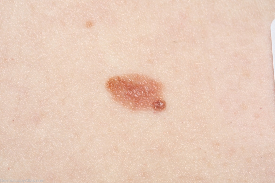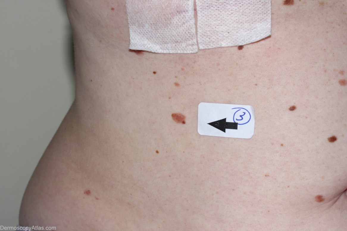

Site: Abdomen
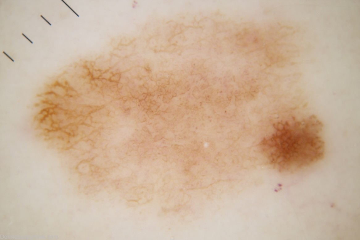
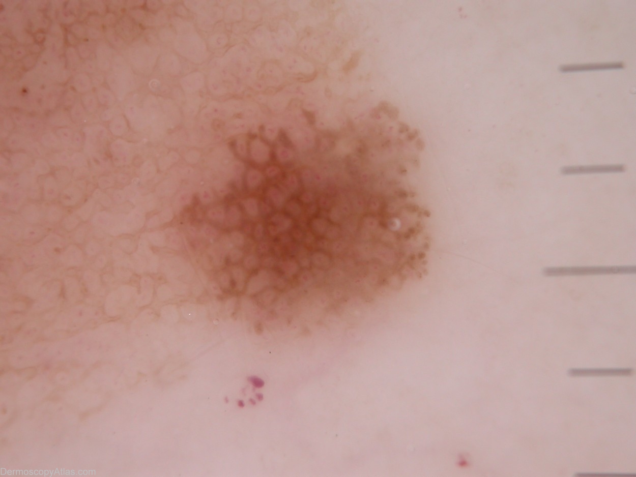
Diagnosis: Nevus dysplastic
Sex: F
Age: 44
Type: Dermlite Non Polarised
Submitted By: Cliff Rosendahl
Description: This naevus has a pattern of lines reticular. In some parts there are bold lines reticular and in other parts there are subtle lines reticular but that is the pattern uniformly throughout the lesion Colour - Eccentric hyperpigmentation of dark brown in a lesion which is otherwise light brown. Clues to melanoma - None.
History:
This 44 year old lady has had 3 previous melanomas. This lesion was noticed on her initial visit to me at which time I removed 2 melanomas (her 2nd and 3rd). This naevus was scheduled for removal a couple of weeks later. The histology report was as follows:-
" Sections show a bland intradermal naevus over the top of which has developed a multifocal junctional dysplastic naevus with mild to moderate atypia. Excision appears complete."
Harald Kittler's assessment prior to seeing the histology report was as follows:-
"Pattern: lines reticular
Colors: eccentric hyperpigmentation
Clues: peripheral brown dots are not a clue to melanoma, because they can be found in any growing lesion. There is a possible pseudopod at the SSE, but whenever I say possible and not striking, I do not count it.
Summary: DDX between Melanoma in situ and Clark's nevus cannot be resolved. With regard to the previous history of multiple melanomas, I would not hesitate to excise the lesion."


