
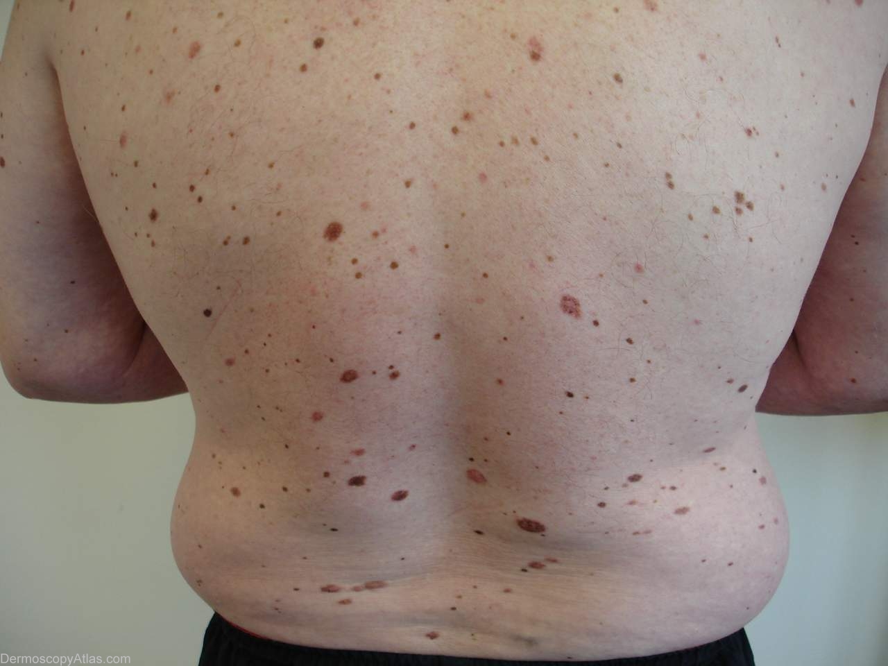
Site: Abdomen
Diagnosis: Nevus dysplastic
Sex: M
Age: 52
Type: Dermlite Polarised
Submitted By: Ian McColl
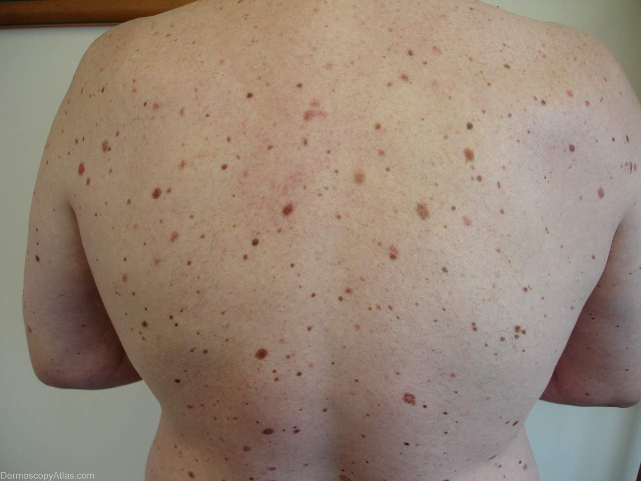
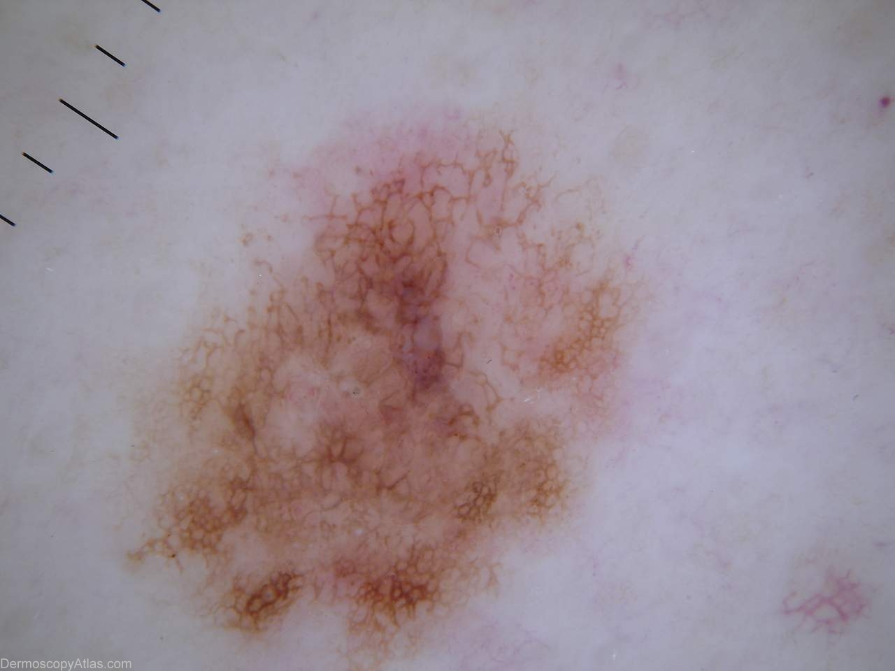
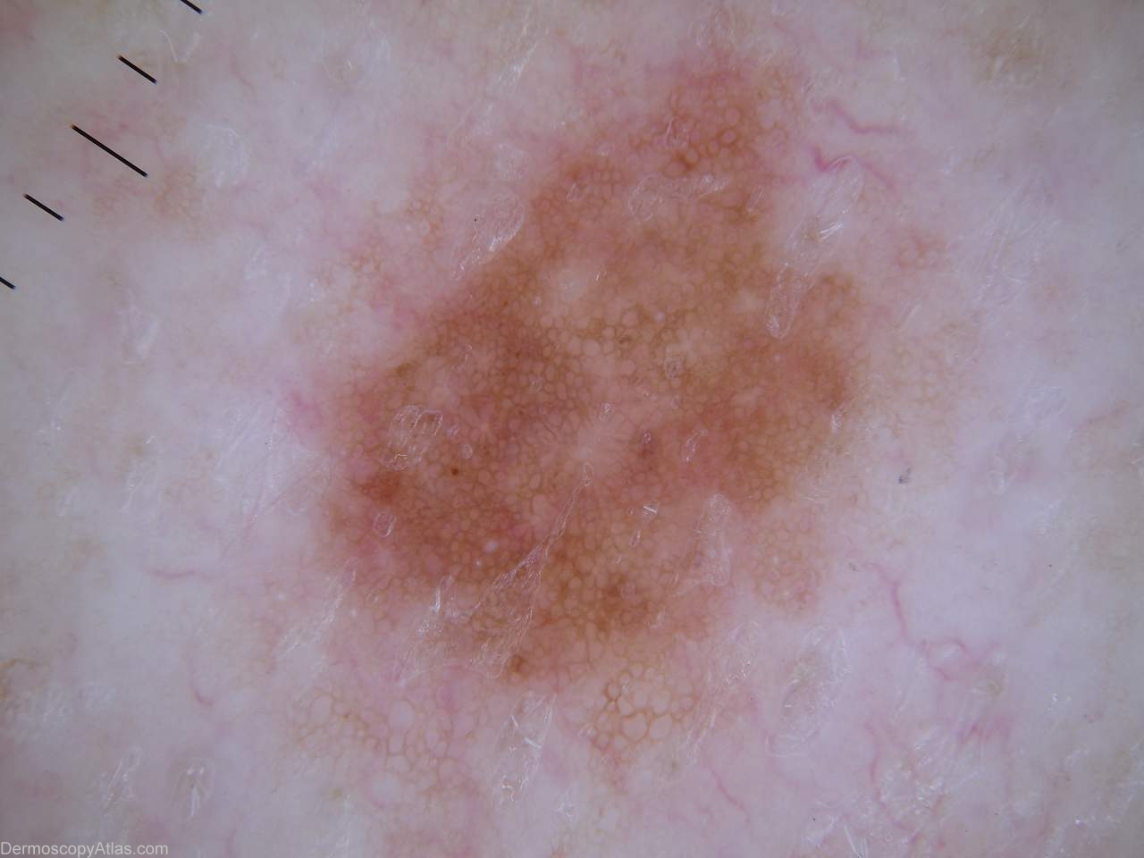
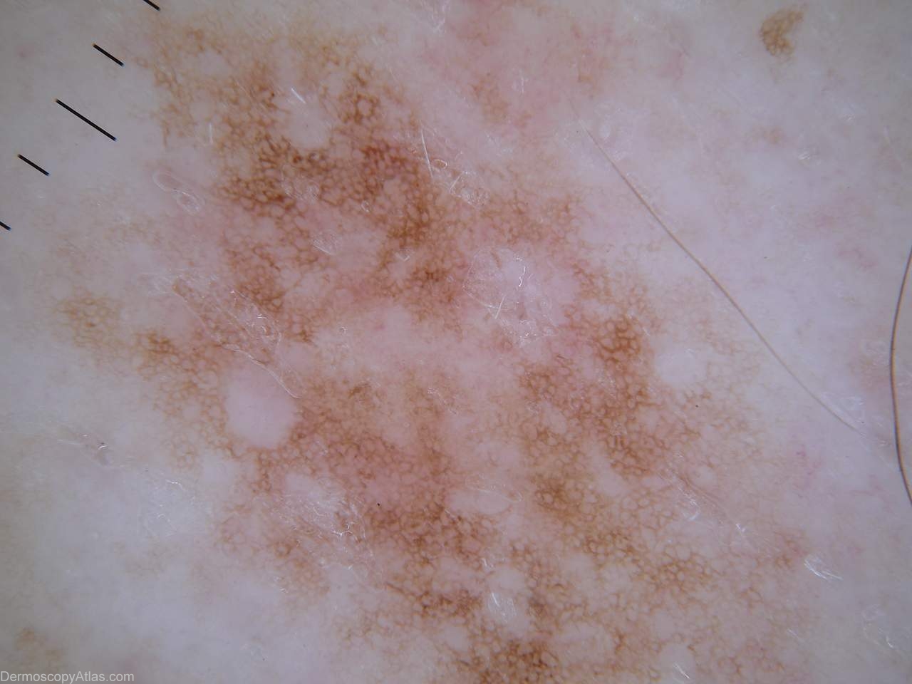
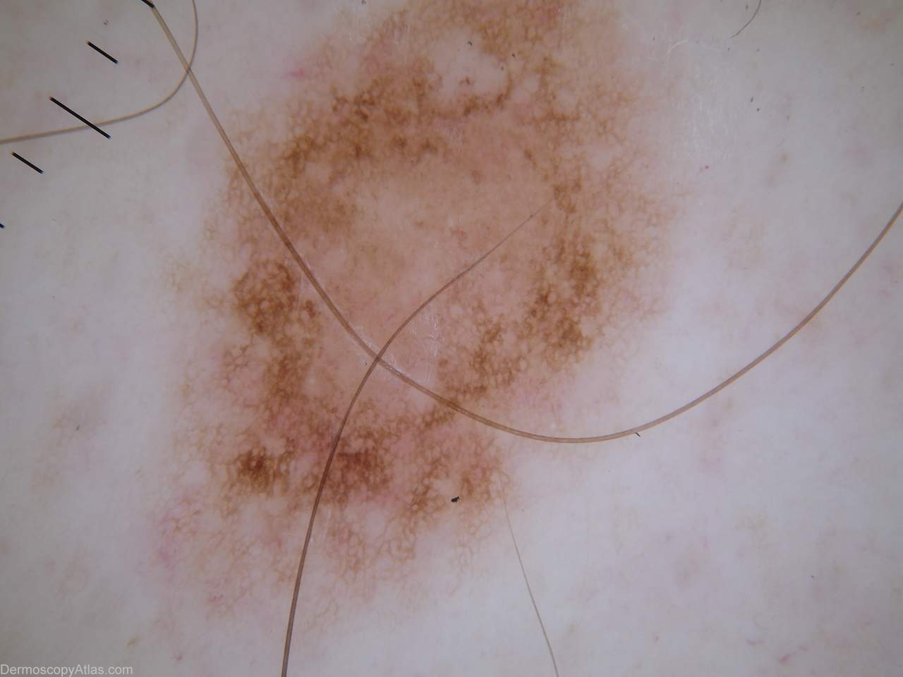
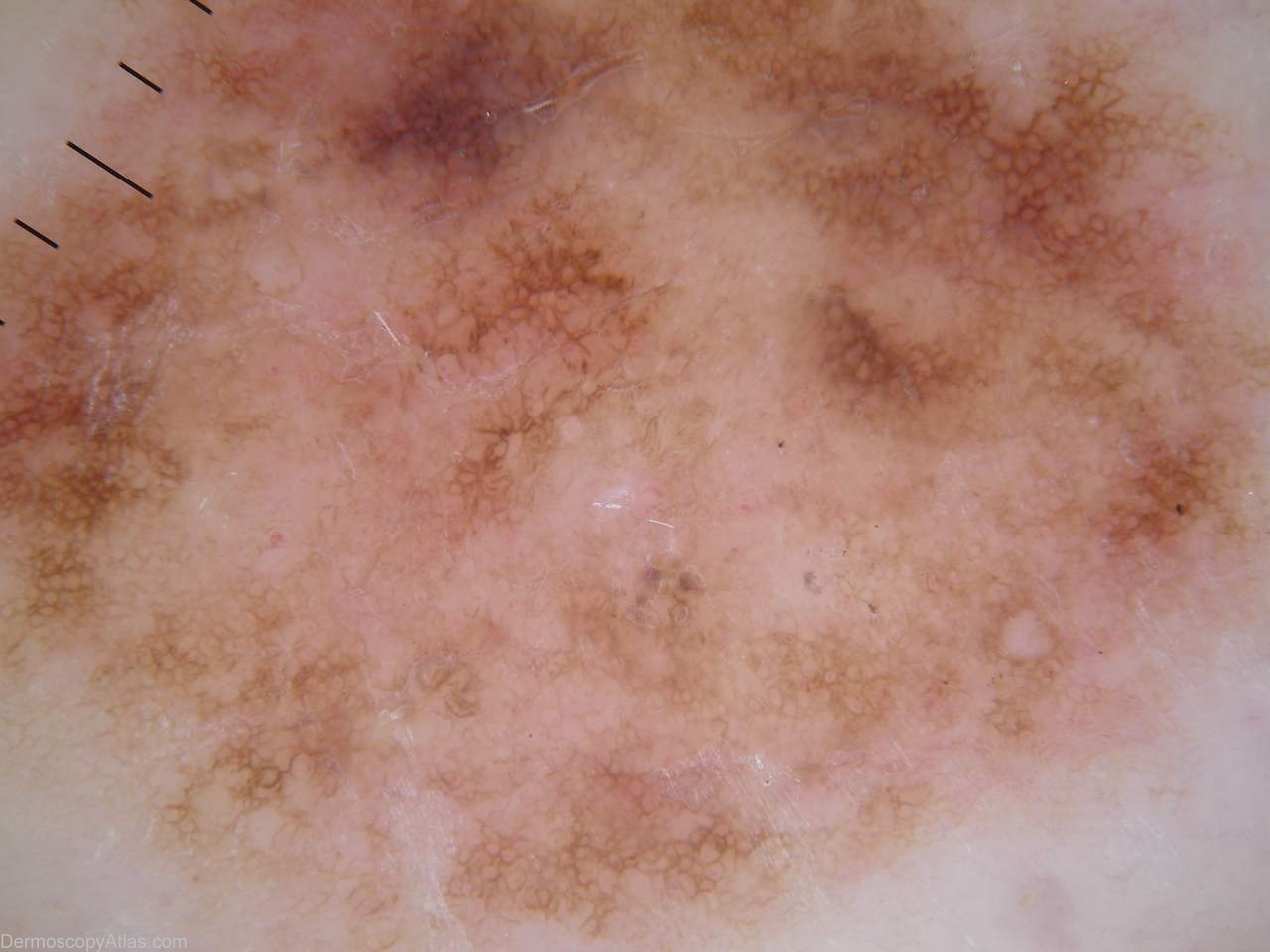
Description: Multiple atypical nevi dysplastic on histology
History:
This man had a melanoma removed more than 10 years ago. He has had total body photography done and has been followed up regularly since but no other melanomas have developed.
These are some examples of his nevi , many of which exhibit a benign atypical nevus pattern. These patterns are described as peripheral network with either hypo or hyperpigmented central areas, peripheral network with a central globular area or a diffuse or patchy generalised network pattern. The more a pattern deviates from those described above the more suspicious the lesion becomes meriting specific monitoring or excision. The greater the architectural disorder the more suspicious the lesion becomes.