
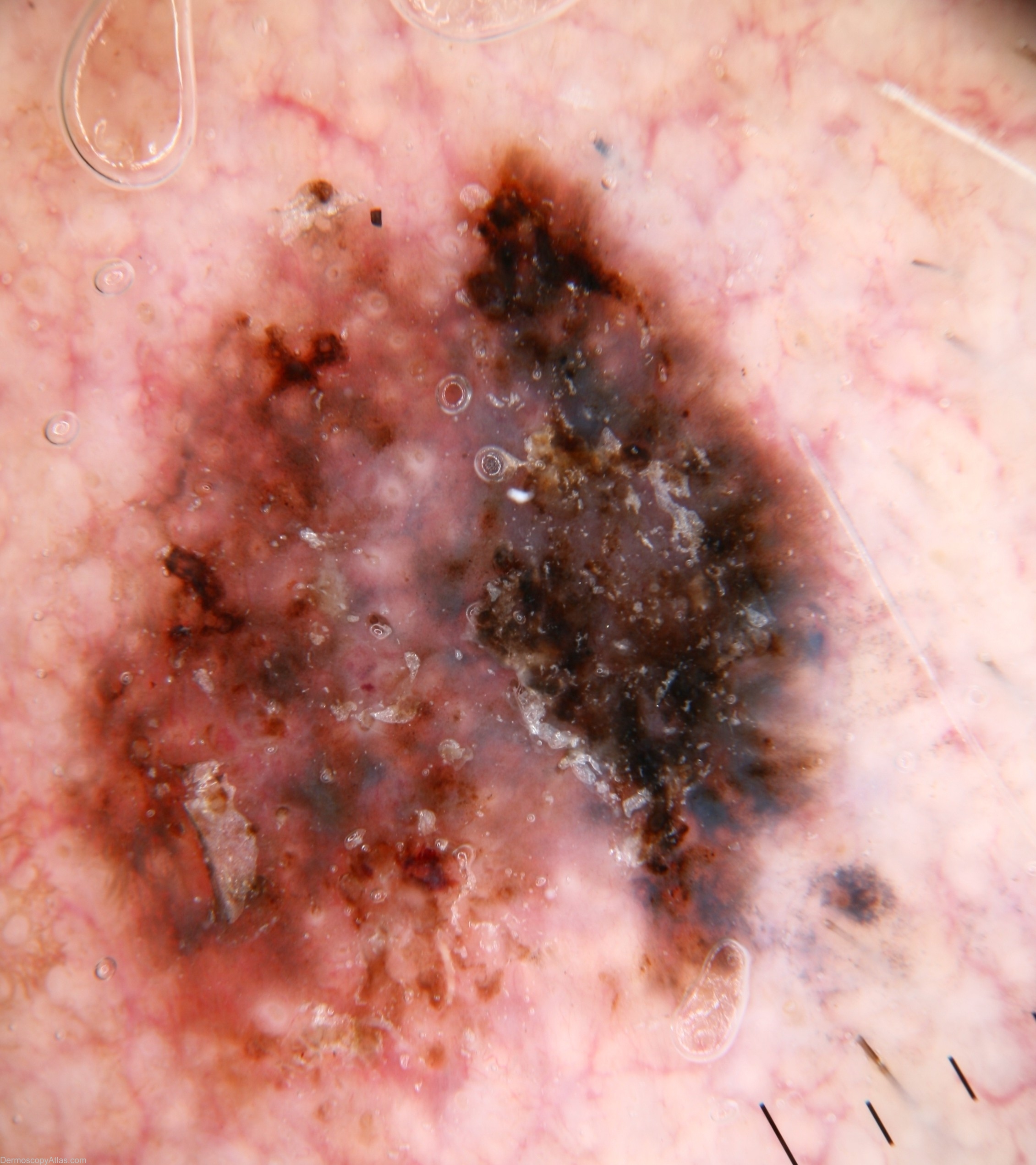
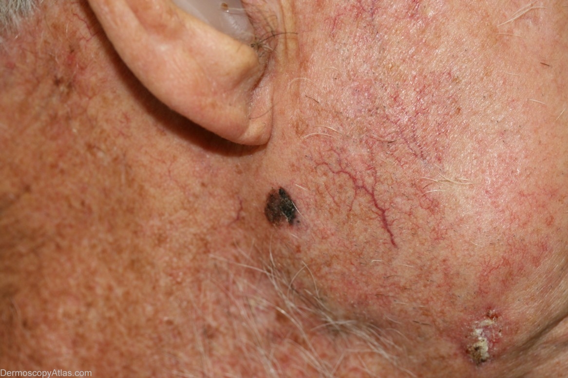
Site: Face
Diagnosis: Melanoma invasive
Sex: M
Age: 85
Type: Dermlite Non Polarised
Submitted By: Cliff Rosendahl
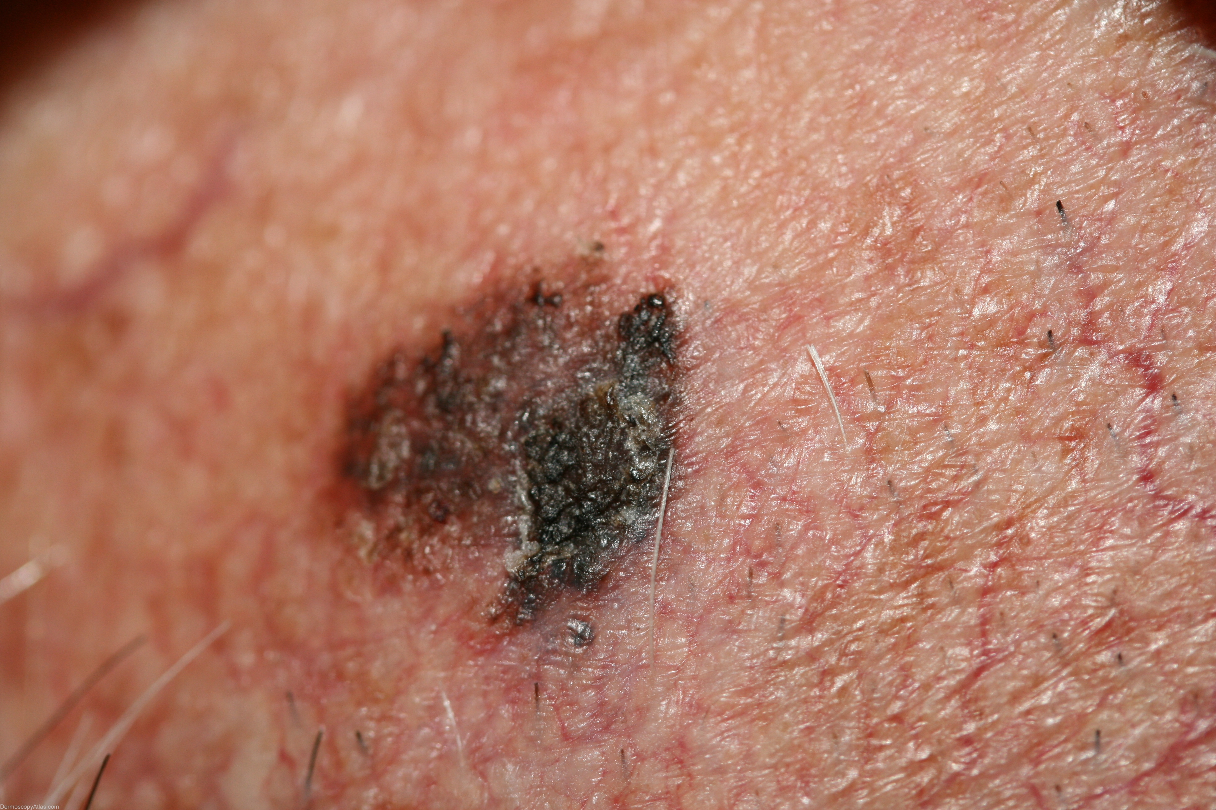
Description: Dermoscopy - This invasive lentigo maligna melanoma ( noted to have some similarities to a map of Australia) has broadened network,broadened network blue-structures, peripheral black globules (clods)and an area of regression separating an "island" from the main body of tumour. It is asymmetrical with several colours.
History:
This lesion was noted macroscopically when the patient presented for another matter.
Histology read:-
(1) Sections show a lentigo maligna melanoma that is Clark level 3. It
is non-ulcerated and has a Breslow thickness of approximately 0.75mm.
Dermal mitoses are present in moderate numbers indicating the lesion is
in vertical growth phase. No lymphovascular or perineural infiltration
is seen. The lesion is completely excised with a minimum clearance of
approximately 6mm.
There was also a BCC which is evident in the clinical image over the lower mandible