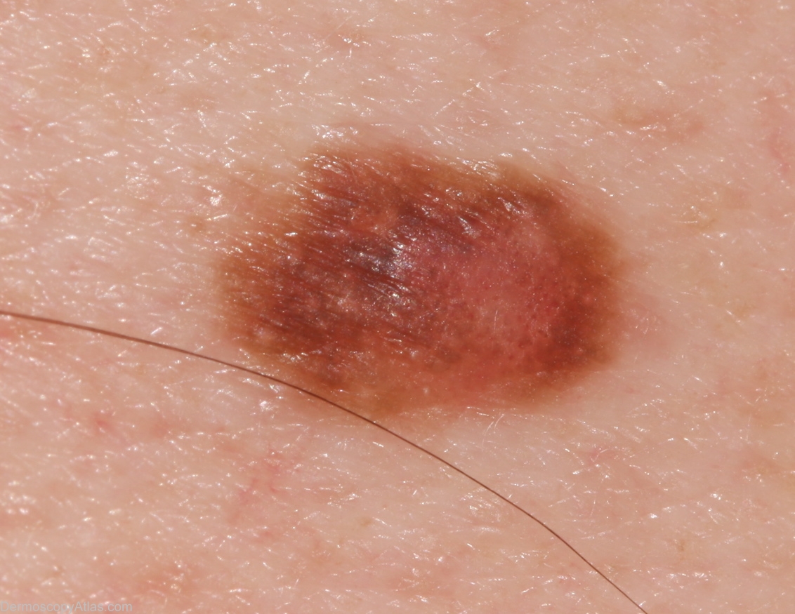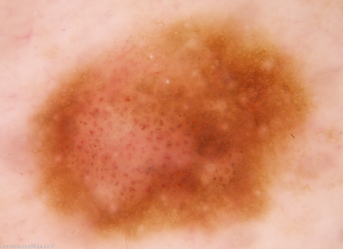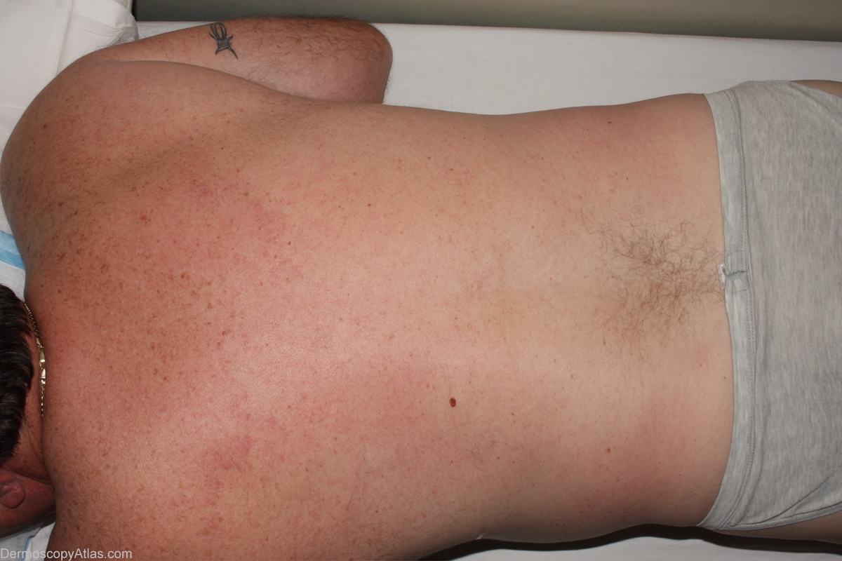

Site: Back
Diagnosis: Nevus dysplastic
Sex: M
Age: 51
Type: Dermlite Non Polarised
Submitted By: Alan Cameron
Description:
History: Solitary lesion on back noted to be enlarging over the previous 6 months by wife. No relevant past or family history. Histology reported as; Sections show a dysplastic naevus of early compound type. There is mild to moderate atypia of junctional melanocytes but good maturation of dermal cells. There is no evidence of malignancy.

