
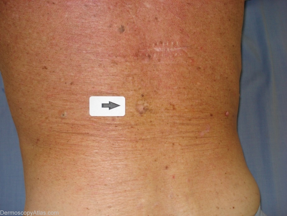
Site: Back
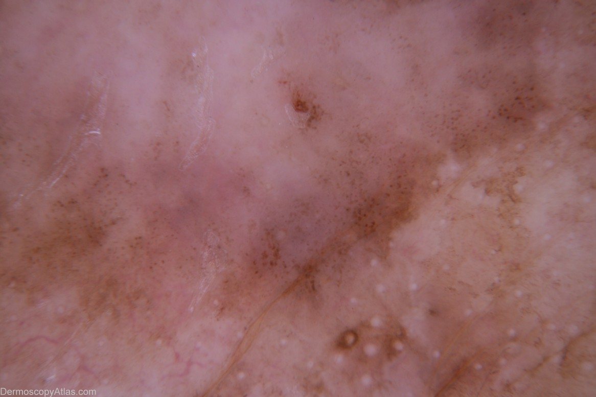
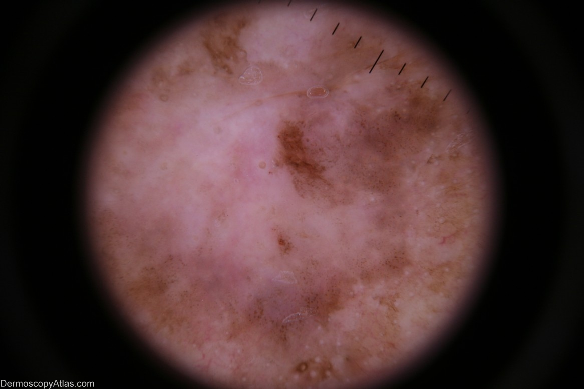
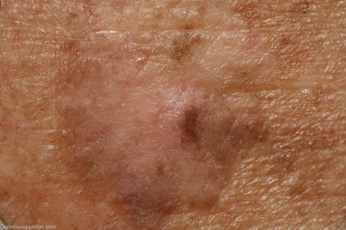
Diagnosis: Pigmented Intraepidermal carcinoma
Sex: M
Age: 68
Type: Dermlite Non Polarised
Submitted By: Cliff Rosendahl
Description: Clinical image
History: This lesion was encountered at a routine skin check at the same time as a level 1 melanoma was discovered on his scalp (image 260 in this atlas). Shave biopsy was reported as "...pigmented intraepidermal carcinoma". The definitive excision was reported as "...pigmented intraepidermal carcinoma and seborrhoeic keratosis". IEC,s do sometimes arise in pre-existing seborrhoeic keratoses. This may be the case here or the two lesions may be separate and in collision. The skin on this man's back was severely sun-damaged.